Copyright
©The Author(s) 2016.
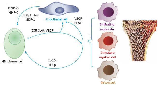
Figure 1 Cell-cell interactions that mediate angiogenesis (in a hypoxic microenvironment).
Interactions between multiple myeloma plasma cells, endothelial cells and different myeloid cells (including infiltrating monocytes, immature myeloid cells such as myeloid-derived suppressor cells, and osteoclasts) stimulate secretion of pro-angiogenic factors. MMP: Matrix metalloproteinase; MM: Multiple myeloma; IL: Interleukin; I-TAC: Interferon-inducible T-cell alpha chemoattractant; SDF-1: Stromal cell-derived factor 1; IGF: Insulin-like growth factor; TGFβ: Transforming growth factor beta; VEGF: Vascular endothelial growth factor; bFGF: Basic fibroblast growth factor.
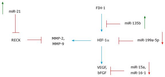
Figure 2 Key mediators of angiogenesis in multiple myeloma and their regulation by miRNAs.
Pro-angiogenic factors are subject to regulation by hypoxia that triggers hypoxia-induce factor 1α, and by other signals (e.g., TGFβ, TNFα), and are fine-tuned by different microRNAs. Green arrows, increased expression levels; red arrows, decreased expression levels. RECK: Reversion-inducing-cysteine-rich protein with kazal motifs; MMP: Matrix metalloproteinase; FIH-1: Factor inhibiting HIF-1; HIF-1α: Hypoxia-inducible factor 1-alpha; VEGF: Vascular endothelial growth factor; bFGF: Basic fibroblast growth factor.
- Citation: Rahat MA, Preis M. Role of microRNA in regulation of myeloma-related angiogenesis and survival. World J Hematol 2016; 5(2): 51-60
- URL: https://www.wjgnet.com/2218-6204/full/v5/i2/51.htm
- DOI: https://dx.doi.org/10.5315/wjh.v5.i2.51


