Copyright
©The Author(s) 2015.
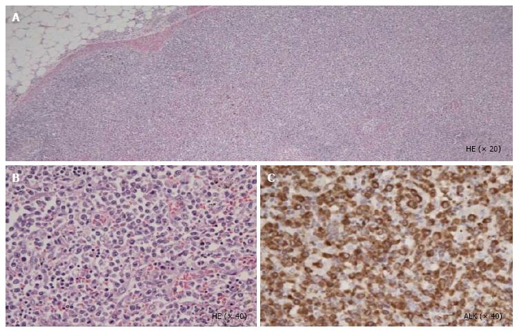
Figure 1 Autopsy was performed.
Hematoxylin-eosin staining in lymph node revealed diffuse proliferation of tumor cells in low power field (A); High power field imaging showed infiltrated atypical small lymphocytes (B); Immunohistochemical staining revealed that the tumor cells expressed anaplastic lymphoma kinase (C). ALK: Anaplastic lymphoma kinase.
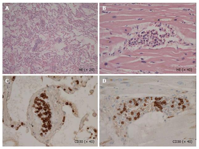
Figure 2 Anaplastic lymphoma kinase positive tumor cells were selectively proliferating in intra lumina of vessels in lung (A, C) and heart (B, D), respectively.
- Citation: Shiroshita K, Kida JI, Matsumoto K, Uemura M, Yamaoka G, Miyai Y, Haba R, Imataki O. Intravascular proliferating anaplastic lymphoma kinase-positive anaplastic large-cell lymphoma. World J Hematol 2015; 4(2): 10-15
- URL: https://www.wjgnet.com/2218-6204/full/v4/i2/10.htm
- DOI: https://dx.doi.org/10.5315/wjh.v4.i2.10


