Copyright
©The Author(s) 2017.
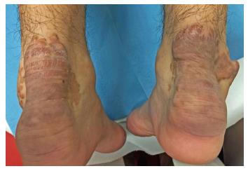
Figure 4 Tendinous xanthomas.
Bilateral Xanthomas of Achilles tendon. Each swelling was localized all over the tendon just above its insertion point to the calcaneal tuberosity. They appear as firm, mobile, painless slowly enlarging subcutaneous nodules which may join together to form a single mass or multilobated masses. They are covered by reddish-brown thickened skin.
- Citation: Mastrolorenzo A, D’Errico A, Pierotti P, Vannucchi M, Giannini S, Fossi F. Pleomorphic cutaneous xanthomas disclosing homozygous familial hypercholesterolemia. World J Dermatol 2017; 6(4): 59-65
- URL: https://www.wjgnet.com/2218-6190/full/v6/i4/59.htm
- DOI: https://dx.doi.org/10.5314/wjd.v6.i4.59


