Copyright
©The Author(s) 2017.
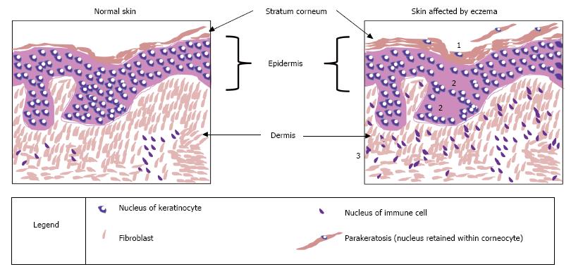
Figure 2 Histological features of atopic eczema.
This figure represents sections of skin biopsies, stained with haematoxylin and eosin, to highlight the changes that can be observed under a light microscope, comparing skin affected by atopic eczema and a control sample. Three characteristic features of atopic eczema are illustrated: Increased thickness of the stratum corneum (hyperkeratosis and parakeratosis) is caused by a disruption to the cornification process; spongiosis occurs when there is a reduction or damage of the proteins involved in tight junctions, thereby leading to uncontrolled movement of fluids in the paracellular space; infiltration of the dermis by immune cells is a sign of the immune response as a primary feature of atopic eczema itself or in response to the entry of allergens through a leaky skin barrier. The effects of eczema: 1: Increased thickness of stratum corneum; 2: Spongiosis - oedema (water retained between cells); 3: Increased number of immune cells.
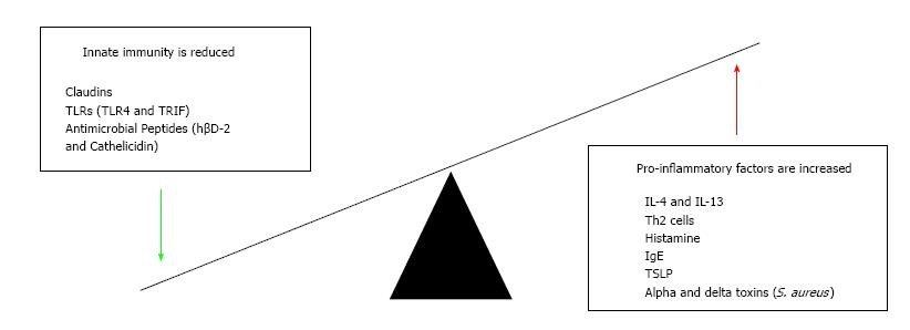
Figure 3 Immune dysfunction in atopic eczema.
This figure summaries the number of immune factors discussed in the review. Increased and decreased levels of different factors contribute to the pathogenesis atopic eczema. TLR: Toll-like receptors; hβD-2: Human beta defensin 2; S. aureus: Staphylococcus aureus; IL: Interleukin; TSLP: Thymic stromal lymphopoietin.
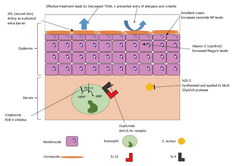
Figure 4 Novel treatment targets in atopic eczema.
This figure highlights the keys roles of treatment: to decrease TEWL, prevent entry of irritants and allergens into the skin and to decrease the action of the immune response. TEWL: Trans-epidermal water loss; IL: Interleukin.
- Citation: Bell DC, Brown SJ. Atopic eczema treatment now and in the future: Targeting the skin barrier and key immune mechanisms in human skin. World J Dermatol 2017; 6(3): 42-51
- URL: https://www.wjgnet.com/2218-6190/full/v6/i3/42.htm
- DOI: https://dx.doi.org/10.5314/wjd.v6.i3.42


