Copyright
©The Author(s) 2016.
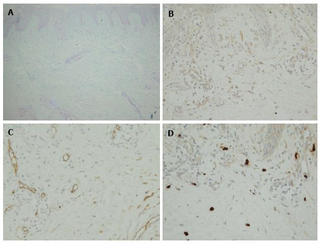
Figure 1 Representative histological findings in active skin lesions of dermatomyositis.
A: Alcian blue (pH 2.5); B: Vimentin; C: CD34; D: Tryptase for mast cell staining.
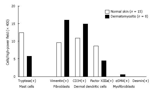
Figure 2 Number of positive cells in active skin lesions of patients with dermatomyositis.
Vimentin+ fibroblasts, CD34+ DDCs and αSMA+ myofibroblasts are increased, but factor XIIIa+ DDCs are decreased in diseased skin compared to normal skin. A significant difference is evident for tryptase-positive mast cells between the diseased and control groups (P < 0.0003). DDCs: Dermal dendritic cells; α-SMA: α-smooth muscle actin.
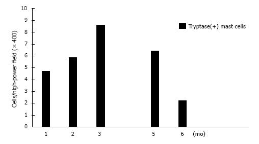
Figure 3 Progress at monthly intervals and mast cell count after occurrence of exanthema.
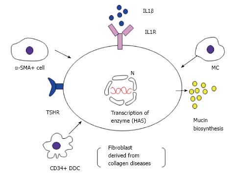
Figure 4 Proposed pathogenic mechanism underlying dermal mucin deposition in dermatomyositis.
α-SMA: α-smooth muscle actin; N: Nucleus; TSHR: Thyroid-stimulating hormone receptor; HAS: Hyaluronan synthase.
- Citation: Yokoyama E, Nakamura Y, Okita T, Nagai N, Muto M. CD34+ dermal dendritic cells and mucin deposition in dermatomyositis. World J Dermatol 2016; 5(2): 65-71
- URL: https://www.wjgnet.com/2218-6190/full/v5/i2/65.htm
- DOI: https://dx.doi.org/10.5314/wjd.v5.i2.65


