Copyright
©The Author(s) 2022.
World J Dermatol. Sep 2, 2022; 10(2): 10-16
Published online Sep 2, 2022. doi: 10.5314/wjd.v10.i2.10
Published online Sep 2, 2022. doi: 10.5314/wjd.v10.i2.10
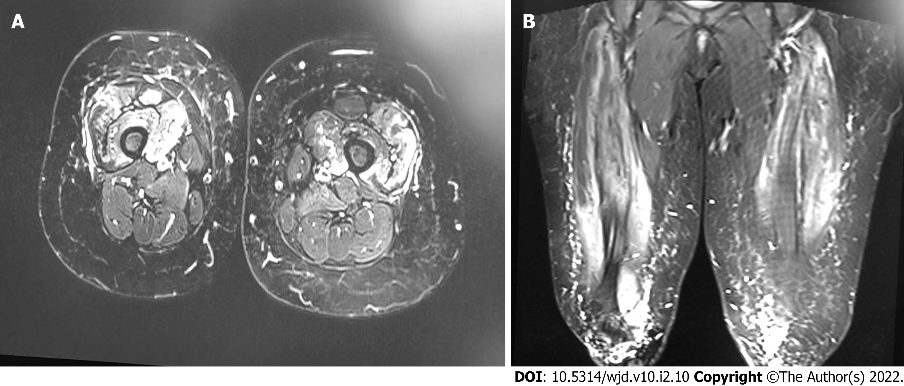
Figure 1 Magnetic resonance imaging scan T1 post-contrast.
A: Axial image of the lower limbs revealed myopathic changes; B: Coronal image shows myopathic changes upon admission.
- Citation: Aly MH, Alshehri AA, Mohammed A, Almaghrabi MA, Alharbi MM. Connection between dermatomyositis and montelukast sodium use: A case report. World J Dermatol 2022; 10(2): 10-16
- URL: https://www.wjgnet.com/2218-6190/full/v10/i2/10.htm
- DOI: https://dx.doi.org/10.5314/wjd.v10.i2.10


