Copyright
©The Author(s) 2020.
World J Anesthesiol. Sep 27, 2020; 9(1): 7-11
Published online Sep 27, 2020. doi: 10.5313/wja.v9.i1.7
Published online Sep 27, 2020. doi: 10.5313/wja.v9.i1.7
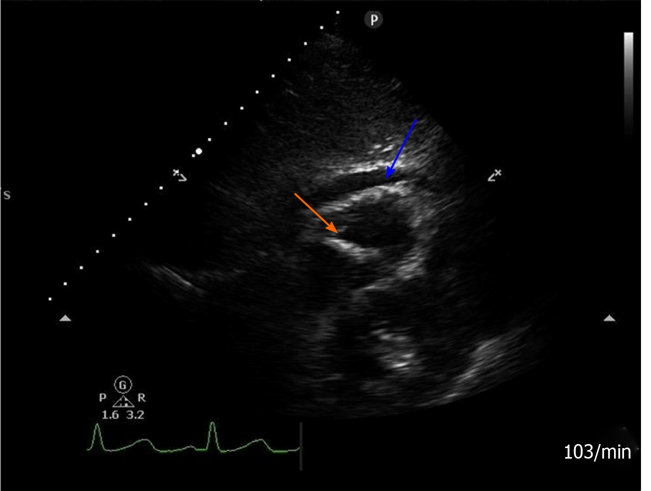
Figure 1 Postoperative transthoracic echocardiography view.
Subxiphoid four chamber view modified for the right ventricle showing a hyperechogenic linear-shaped image attached to the apical portion of the right ventricle (orange arrow), and pericardial effusion (blue arrow).
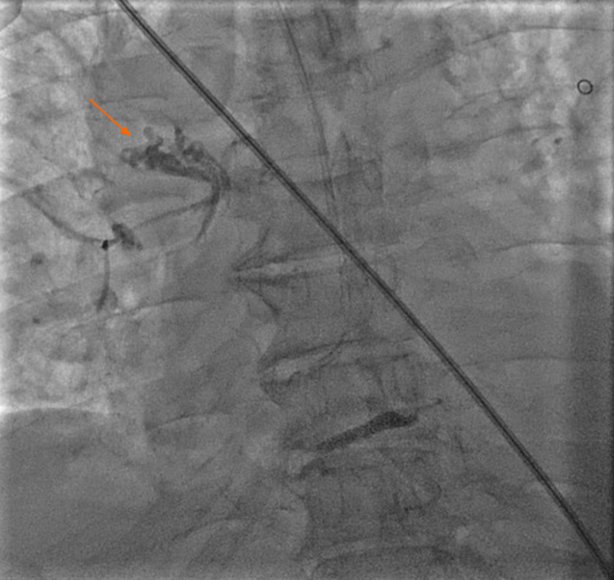
Figure 2 Postoperative coronary angiography examination.
Coronary angiography showed an opaque lesion on the right pulmonary artery (orange arrow).
- Citation: Xu ZZ, Li HJ, Li X, Zhang H. Cement-related embolism after lumbar vertebroplasty: A case report. World J Anesthesiol 2020; 9(1): 7-11
- URL: https://www.wjgnet.com/2218-6182/full/v9/i1/7.htm
- DOI: https://dx.doi.org/10.5313/wja.v9.i1.7


