Copyright
©The Author(s) 2015.
World J Anesthesiol. Nov 27, 2015; 4(3): 49-57
Published online Nov 27, 2015. doi: 10.5313/wja.v4.i3.49
Published online Nov 27, 2015. doi: 10.5313/wja.v4.i3.49
Figure 1 Lumbar medial branch anatomy.
Left anterior oblique illustration (L3 to S1): Spinous processes. mal: Maillo-accessory ligament; nr: Nerve root; I: Inferior articular process; S: Superior articular process; sb: Superior branch from medial branch; ib: Inferior branch of medial branch; dr: Dorsal ramus; mb: Medial branch[13,14].
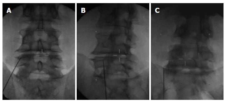
Figure 3 Different views of an electrode placed for an L4 medial branch neurotomy.
A: Antero-posterior view; B: Corresponding oblique view; C: Antero-posterior view of an electrode placed for an L5 medial branch neurotomy[14].
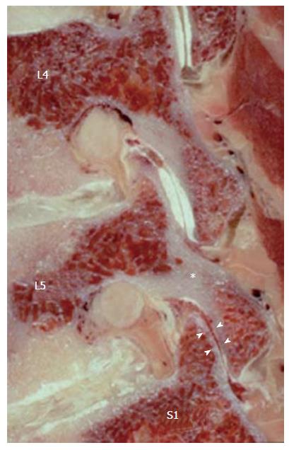
Figure 4 Sagittal section through the neuroforamina of a severely degenerated lower lumbar spine of a 70-year-old man.
The z-joints are in a subluxated position due to the loss of segmental height. The pars interarticularis of L5 is being eroded superiorly by the inferior articular process of L4 and inferiorly by the superior articular process of S1 (*). Such pars erosion is a prerequisite for the development of degenerative spondylolisthesis. There is no cartilage in the L5/S1 z-joint (arrow heads). Z-joints: Zygapophysial joints.
- Citation: Klessinger S. Zygapophysial joint pain in selected patients. World J Anesthesiol 2015; 4(3): 49-57
- URL: https://www.wjgnet.com/2218-6182/full/v4/i3/49.htm
- DOI: https://dx.doi.org/10.5313/wja.v4.i3.49


