Copyright
©2014 Baishideng Publishing Group Inc.
World J Orthop. Nov 18, 2014; 5(5): 574-584
Published online Nov 18, 2014. doi: 10.5312/wjo.v5.i5.574
Published online Nov 18, 2014. doi: 10.5312/wjo.v5.i5.574
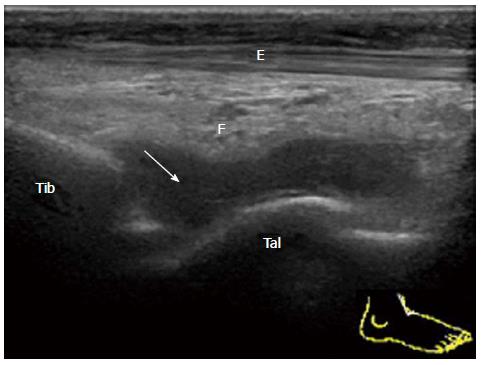
Figure 2 Severe tibiotalar joint synovitis in rheumatoid arthritis.
Sagittal grey-scale sonogram of the anterior recess of the tibiotalar joint shows that a normal hyperechoic anterior fat pad (F) is displaced anteriorly by hypoechoic synovium (arrow). Tib: Tibia; Tal: Talus; F: Fat pad; E: Extensor hallucis longus.
- Citation: Suzuki T. Power Doppler ultrasonographic assessment of the ankle in patients with inflammatory rheumatic diseases. World J Orthop 2014; 5(5): 574-584
- URL: https://www.wjgnet.com/2218-5836/full/v5/i5/574.htm
- DOI: https://dx.doi.org/10.5312/wjo.v5.i5.574


