Copyright
©The Author(s) 2018.
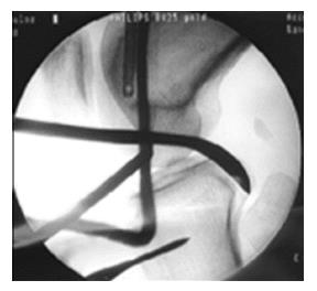
Figure 1 Tibial tunnel opening under image intensifier.
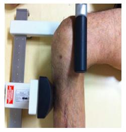
Figure 2 Proper position of tibia and femur for Telos Stress Device.
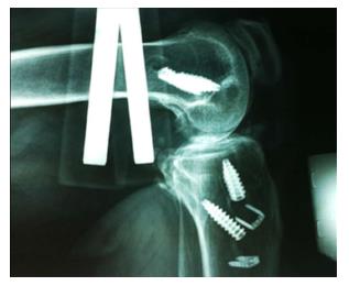
Figure 3 Anteroposterior force on tibia through Telos Device leading to posterior translation.
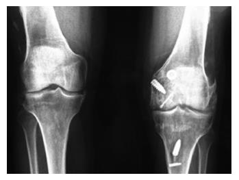
Figure 4 Anteroposterior knee x-rays for evaluation of arthritis progression (Kellgren and Lawrence grade 3).
- Citation: Gliatis J, Anagnostou K, Tsoumpos P, Billis E, Papandreou M, Plessas S. Complex knee injuries treated in acute phase: Long-term results using Ligament Augmentation and Reconstruction System artificial ligament. World J Orthop 2018; 9(3): 24-34
- URL: https://www.wjgnet.com/2218-5836/full/v9/i3/24.htm
- DOI: https://dx.doi.org/10.5312/wjo.v9.i3.24


