Copyright
©The Author(s) 2018.
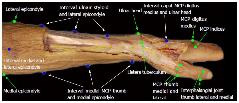
Figure 1 Landmarks used to outline the arm.
MCP: Metacarpo phalangeal.
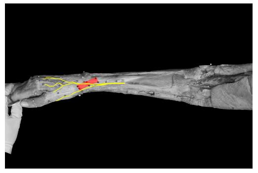
Figure 2 Course of the superficial branch of the radial nerve compared to the first extensor compartment.
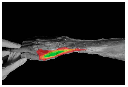
Figure 3 “Safe zone” gradient from red (95% nerve density) to green (0% nerve density).
Figure 4 Distance between first and second branch of the superficial branch of the radial nerve for all 20 arms.
SBRN: Superficial branch of the radial nerve.
Figure 5 Course of 20 superficial branch of the radial nerve (Yellow) and 20 lateral antebrachial cutaneous nerve (Green) compared to the first extensor compartment.
- Citation: Poublon AR, Kleinrensink GJ, Kerver AL, Coert JH, Walbeehm ET. Optimal surgical approach for the treatment of Quervains disease: A surgical-anatomical study. World J Orthop 2018; 9(2): 7-13
- URL: https://www.wjgnet.com/2218-5836/full/v9/i2/7.htm
- DOI: https://dx.doi.org/10.5312/wjo.v9.i2.7


