Copyright
©The Author(s) 2017.
World J Orthop. Jul 18, 2017; 8(7): 567-573
Published online Jul 18, 2017. doi: 10.5312/wjo.v8.i7.567
Published online Jul 18, 2017. doi: 10.5312/wjo.v8.i7.567
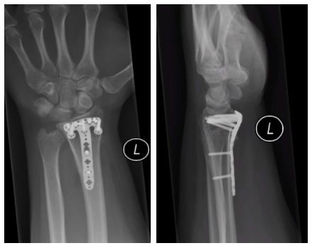
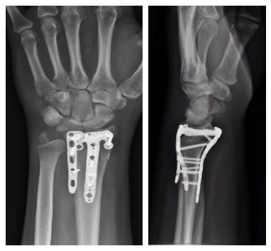
Figure 1 Posteroanterior and lateral radiographs of fracture fixation using a Synthes variable angle 2.
4 distal radius rim plate to highlight the distal placement of the implant.
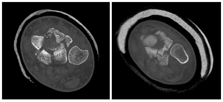
Figure 2 Axial computerised tomograms showing far distal involvement and comminution of the distal radio-ulnar joint.
Figure 3 Axial computerised tomograms showing far distal involvement and comminution of the distal radio-ulnar joint.
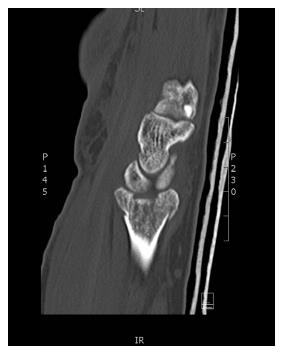
Figure 4 Sagittal computerized tomogram showing the pattern of significant intra-articular comminution of the far distal dorso-ulnar and volar-ulnar regions of the distal radius and the volar lip marginal fragment.
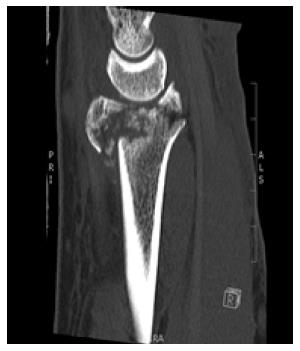
Figure 5 PA and lateral radiograph showing combined dorsal and volar plating using a volar rim plate.
- Citation: Spiteri M, Roberts D, Ng W, Matthews J, Power D. Distal radius volar rim plate: Technical and radiographic considerations. World J Orthop 2017; 8(7): 567-573
- URL: https://www.wjgnet.com/2218-5836/full/v8/i7/567.htm
- DOI: https://dx.doi.org/10.5312/wjo.v8.i7.567


