Copyright
©The Author(s) 2017.
World J Orthop. Mar 18, 2017; 8(3): 229-241
Published online Mar 18, 2017. doi: 10.5312/wjo.v8.i3.229
Published online Mar 18, 2017. doi: 10.5312/wjo.v8.i3.229
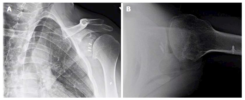
Figure 1 Anteroposterior (A) and lateral (B) X-rays of a 39-year-old male with dislocation arthropathy status post instability procedure with metal anchors.
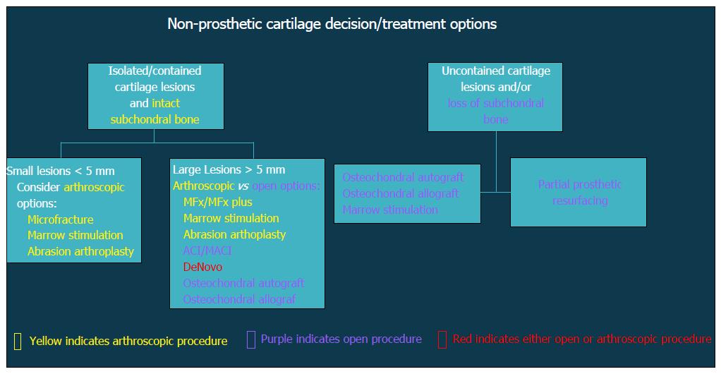
Figure 2 Flow-chart demonstrating decision algorithm for non-prosthetic cartilage treatment options.
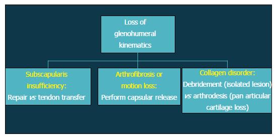
Figure 3 Flow-chart depicting treatment options after loss of glenohumeral kinematics.
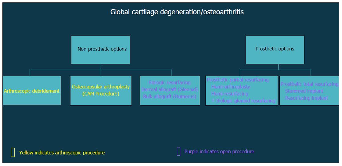
Figure 4 Flow-chart demonstrating decision algorithm for non-prosthetic vs prosthetic treatment options that include arthroscopic and open procedures.
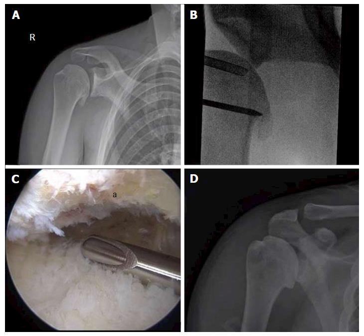
Figure 5 Comprehensive arthroscopy management of glenohumeral arthropathy.
A: Images from a 37-year-old male with instability arthropathy demonstrating preoperative anteroposterior radiograph with large inferior humeral head osteophyte and loss of glenohumeral joint space; B: Intra-operative fluoroscopy localization of extent of inferior humeral head osteophyte; C: Intra-operative arthroscopic image viewing from posterior portal, demonstrating inferior humeral neck (a) status post debridement of osteophyte, the arthroscopic shaver is on the inferior capsule; D: Post-operative anteroposterior radiograph demonstrating debridement of osteophyte and biceps tenodesis with a biocomposite screw.
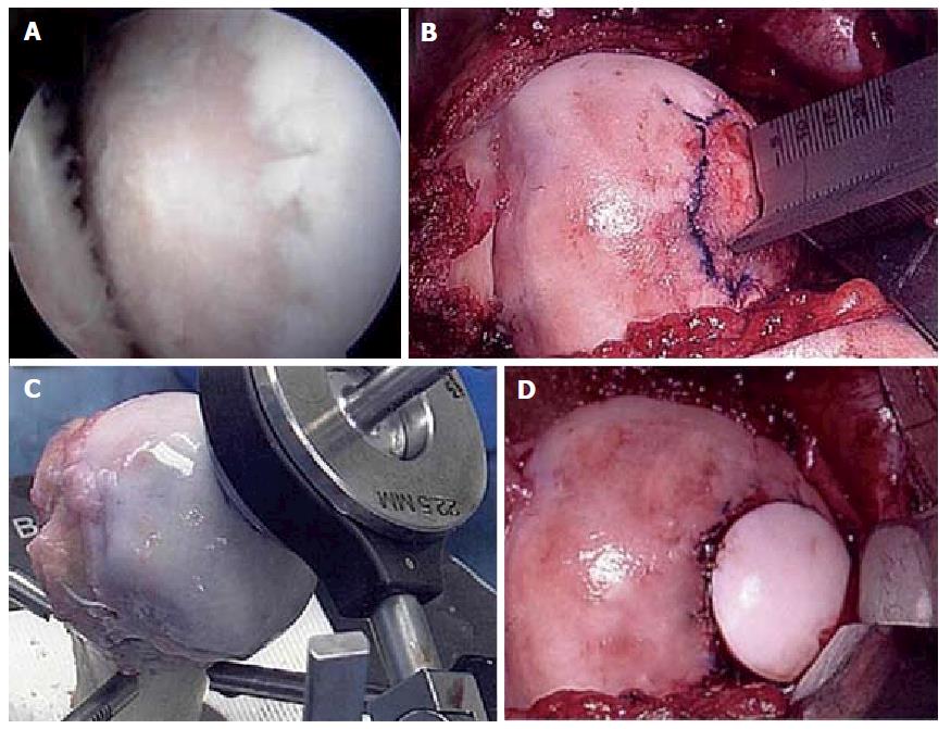
Figure 6 Fresh osteochondral allograft transplantation.
A: Intra-operative arthroscopic image of central humeral articular lesion while viewing from a posterior portal in a 39-year-old patient; B: After an open approach, preparation of the central lesion; C: Harvesting a corresponding osteochondral plug from a size-matched, fresh allograft humerus; D: Status post insertion of the osteochondral plug into the defect.
- Citation: Waterman BR, Kilcoyne KG, Parada SA, Eichinger JK. Prevention and management of post-instability glenohumeral arthropathy. World J Orthop 2017; 8(3): 229-241
- URL: https://www.wjgnet.com/2218-5836/full/v8/i3/229.htm
- DOI: https://dx.doi.org/10.5312/wjo.v8.i3.229


