Copyright
©The Author(s) 2017.
World J Orthop. Feb 18, 2017; 8(2): 208-211
Published online Feb 18, 2017. doi: 10.5312/wjo.v8.i2.208
Published online Feb 18, 2017. doi: 10.5312/wjo.v8.i2.208
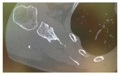
Figure 1 Axial computed tomography showing defect of scapular body.
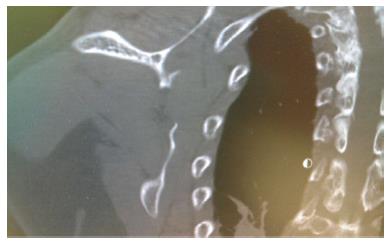
Figure 2 Coronal computed tomography showing defect of scapular body.
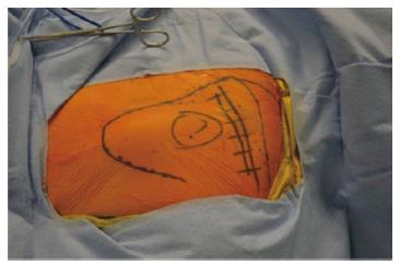
Figure 3 Borders of scapula, scapular spine, incision site (dashed line) and area over defect were marked.
Figure 4 The 10 cm incision made just inferior to scapula spine, running from medial to lateral.
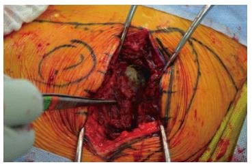
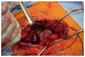
Figure 5 The 4 cm × 2 cm scapular defect exposed.
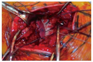
Figure 6 The dermal jacket is sewn into the defect.
- Citation: Grau L, Chen K, Alhandi AA, Goldberg B. Novel technique for a symptomatic subscapularis herniation through a scapular defect. World J Orthop 2017; 8(2): 208-211
- URL: https://www.wjgnet.com/2218-5836/full/v8/i2/208.htm
- DOI: https://dx.doi.org/10.5312/wjo.v8.i2.208


