Copyright
©The Author(s) 2017.
World J Orthop. Feb 18, 2017; 8(2): 142-148
Published online Feb 18, 2017. doi: 10.5312/wjo.v8.i2.142
Published online Feb 18, 2017. doi: 10.5312/wjo.v8.i2.142
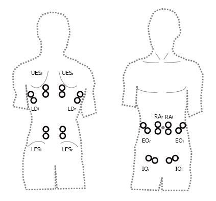
Figure 1 12-lead EMG trunk-setup.
Single muscles: RAri/le: M. rec. abd. right/left; EOri/le: M. obl. ext. abd. right/left; IOri/le: M. obl. int. abd. right/left; LDri/le: M. latis. dorsi right/left; UESri/le: M. erec. spinae thoracic (T9) right/left; LESri/le: M. erec. spinae lumbar (L3) right/left.
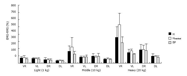
Figure 2 Neuromuscular activity (EMG-RMS; %) of trunk areas for healthy controls (H; Hmatched) and back pain patients for the lifting tasks with 1, 10 and 20 kg (VR/VL: RA, EO, IOri/le; DR/DL: LD, UES, LESri/le).
BPP: Back pain patients; VR/VL: Right/left ventral area; DR/DL: Right/left dorsal area.
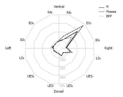
Figure 3 Polarplot of neuromuscular activity (EMG-RMS; %) of the 12 trunk muscles in healthy controls (H/Hmatched) and back pain patients for lifting of middle load (10 kg).
- Citation: Mueller J, Engel T, Kopinski S, Mayer F, Mueller S. Neuromuscular trunk activation patterns in back pain patients during one-handed lifting. World J Orthop 2017; 8(2): 142-148
- URL: https://www.wjgnet.com/2218-5836/full/v8/i2/142.htm
- DOI: https://dx.doi.org/10.5312/wjo.v8.i2.142


