Copyright
©The Author(s) 2017.
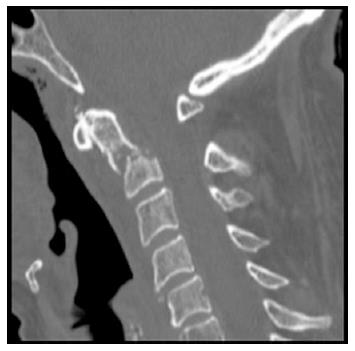
Figure 1 Computed tomography-scan at admission to the hospital showing a transverse fracture line above C2 vertebral body and below transverse ligament of the odontoid process (type II fracture).
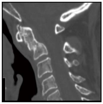
Figure 2 Interval computed tomography-scan 6 mo after the index injury.
No healing is demonstrated, sclerotic bone margins are demonstrated at the fracture fragments.
Figure 3 Computed tomography-scan performed at the end of 3 mo of anabolic therapy with Teriparatide confirming a complete fusion of the fracture with acceptable alignment.
- Citation: Pola E, Pambianco V, Colangelo D, Formica VM, Autore G, Nasto LA. Teriparatide anabolic therapy as potential treatment of type II dens non-union fractures. World J Orthop 2017; 8(1): 82-86
- URL: https://www.wjgnet.com/2218-5836/full/v8/i1/82.htm
- DOI: https://dx.doi.org/10.5312/wjo.v8.i1.82


