Copyright
©The Author(s) 2016.
World J Orthop. Sep 18, 2016; 7(9): 623-627
Published online Sep 18, 2016. doi: 10.5312/wjo.v7.i9.623
Published online Sep 18, 2016. doi: 10.5312/wjo.v7.i9.623
Figure 1 Initial trauma screening revealed a hemodynamically stable patient with acomplicated (Gustilo type 3) subtalar joint dislocation of the right leg.
A and B: Gustilo grade 3 complicated lateral dislocation of the right foot; C: Radiograph showing lateral dislocation of the subtalar joint.
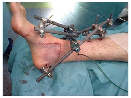
Figure 2 The wound is closed primarily after meticulous debridement and an external fixator is applied.
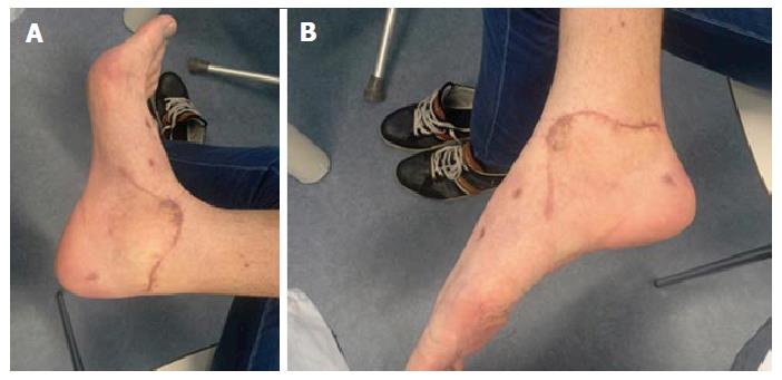
Figure 3 Excellent ankle function is restored at final follow-up (A and B).
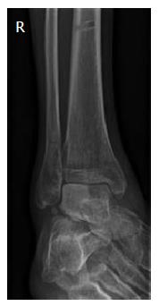
Figure 4 Radiograph of the ankle at follow-up, showing early signs of avascular talar necrosis.
- Citation: Veltman ES, Steller EJ, Wittich P, Keizer J. Lateral subtalar dislocation: Case report and review of the literature. World J Orthop 2016; 7(9): 623-627
- URL: https://www.wjgnet.com/2218-5836/full/v7/i9/623.htm
- DOI: https://dx.doi.org/10.5312/wjo.v7.i9.623


