Copyright
©The Author(s) 2016.
World J Orthop. May 18, 2016; 7(5): 301-307
Published online May 18, 2016. doi: 10.5312/wjo.v7.i5.301
Published online May 18, 2016. doi: 10.5312/wjo.v7.i5.301
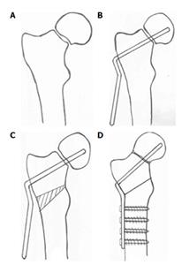
Figure 1 Diagram showing stages of valgus osteotomy.
A: Closed reduction; B: Insertion of blade plate device; C: Excision of lateral wedge; D: Final correction after plate fixation.
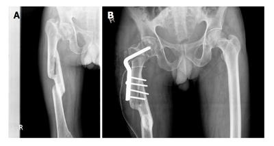
Figure 2 Problems with neglected fractures.
A: Anteroposterior radiograph of pelvis of a 41-year-old male with a 3-mo-old nonunion neck of femur fracture and associated malunited femur fracture; B: At 4-year follow-up showing union, when the Harris hip score was 88.
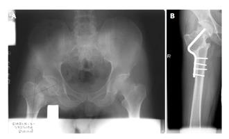
Figure 3 Problems with excess valgus.
A: Anteroposterior radiograph of pelvis of a 33-year-old male with a 3-mo-old nonunion neck of femur fracture; B: At 10-year follow-up, showing excess valgus, when the Harris hip score was 68.
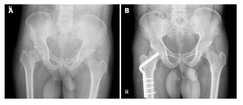
Figure 4 Ideal valgus correction.
A: Anteroposterior radiograph of pelvis of a 45-year-old male with a 1-mo-old nonunion neck of femur fracture; B: At 5-year follow-up showing similar valgus orientation as the opposite hip, when the Harris hip score was 85.
- Citation: Varghese VD, Livingston A, Boopalan PR, Jepegnanam TS. Valgus osteotomy for nonunion and neglected neck of femur fractures. World J Orthop 2016; 7(5): 301-307
- URL: https://www.wjgnet.com/2218-5836/full/v7/i5/301.htm
- DOI: https://dx.doi.org/10.5312/wjo.v7.i5.301


