Copyright
©The Author(s) 2016.
World J Orthop. Mar 18, 2016; 7(3): 188-194
Published online Mar 18, 2016. doi: 10.5312/wjo.v7.i3.188
Published online Mar 18, 2016. doi: 10.5312/wjo.v7.i3.188
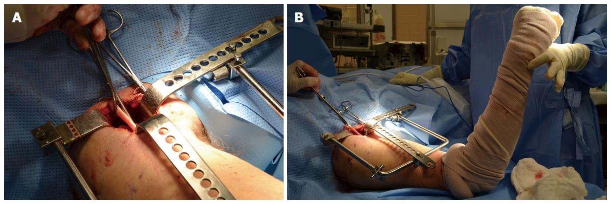
Figure 1 Intraoperative image.
A: After incision of the proximal 1 cm insertion of the pectoralis tendon, the retracted and scarred long head of the biceps tendon is brought into the wound; B: Long head of the biceps tendon tenodesis is performed at 60° of elbow flexion.
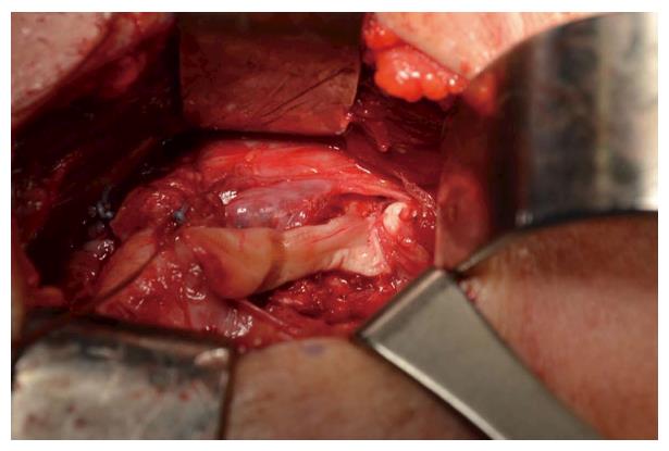
Figure 2 Intraoperative image of the biceps tenodesis: Suprapectoralis tenodesis at the bottom of bicipital groove using 7 mm × 23 mm bioabsorbable interference screw (Milagro, DePuy Mitek, MA, United States).
- Citation: McMahon PJ, Speziali A. Outcomes of tenodesis of the long head of the biceps tendon more than three months after rupture. World J Orthop 2016; 7(3): 188-194
- URL: https://www.wjgnet.com/2218-5836/full/v7/i3/188.htm
- DOI: https://dx.doi.org/10.5312/wjo.v7.i3.188


