Copyright
©The Author(s) 2016.
World J Orthop. Oct 18, 2016; 7(10): 650-656
Published online Oct 18, 2016. doi: 10.5312/wjo.v7.i10.650
Published online Oct 18, 2016. doi: 10.5312/wjo.v7.i10.650
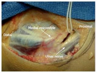
Figure 1 Ulnar nerve anatomy at the elbow.
The ulnar nerve courses posterior to the intermuscular septum and adjacent to the triceps. It passes posterior to the medial epicondyle before entering into the cubital tunnel.
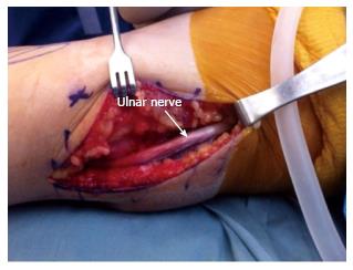
Figure 2 Dissection of the ulnar nerve.
The ulnar nerve is identified proximal to the cubital tunnel and posterior to the medial intermuscular septum. Dissection of the nerve begins proximally at the arcade of Struthers and is continued distally to the two heads of the flexor carpi ulnaris muscle.
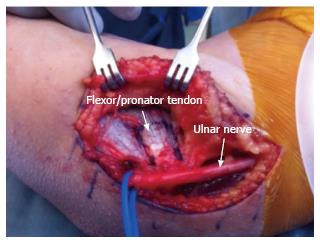
Figure 3 Protection of ulnar nerve.
The surgeon must be aware of the anatomy of the ulnar nerve and protect it until the ulnar collateral ligament reconstruction is complete.
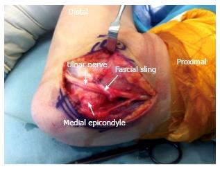
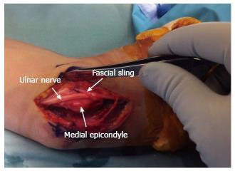
Figure 4 Anterior subcutaneous transposition of the ulnar nerve in flexion.
A band of medial intermuscular septum is used as a sling to hold the nerve in place. One end of the band is excised beginning 8 cm proximal to the medial epicondyle, and the other end is left attached to the medial epicondyle. The strip is sutured onto the fascia overlying the flexor-pronator musculature.
Figure 5 Anterior subcutaneous transposition of the ulnar nerve in extension.
The inverted V-shaped sling prevents the nerve from falling behind the medial epicondyle.
- Citation: Conti MS, Camp CL, Elattrache NS, Altchek DW, Dines JS. Treatment of the ulnar nerve for overhead throwing athletes undergoing ulnar collateral ligament reconstruction. World J Orthop 2016; 7(10): 650-656
- URL: https://www.wjgnet.com/2218-5836/full/v7/i10/650.htm
- DOI: https://dx.doi.org/10.5312/wjo.v7.i10.650


