Copyright
©The Author(s) 2016.
World J Orthop. Oct 18, 2016; 7(10): 628-637
Published online Oct 18, 2016. doi: 10.5312/wjo.v7.i10.628
Published online Oct 18, 2016. doi: 10.5312/wjo.v7.i10.628
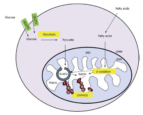
Figure 1 Cellular energy production.
In skeletal muscle, glucose enters the cell through glucose transporter type 1 or 4 (GLUT1 or GLUT4, respectively). Glucose is converted to pyruvate in the glycolysis pathway. Pyruvate is transported across the outer and inner mitochondrial membranes (OMM and IMM, respectively) and into the mitochondrial matrix where it is converted into acetyl-coA. Fatty acids undergo β-oxidation in the mitochondria, creating acetyl-coA and NADH. Acetyl-coA is utilized by the Kreb’s cycle, creating NADH and succinate which enter the electron transport chain (ETC) at complex I and II, respectively. The movement of electrons through the ETC is coupled to the production of ATP in a process called OXPHOS. IMS: Intermembrane space; OXPHOS: Oxidative phosphorylation.
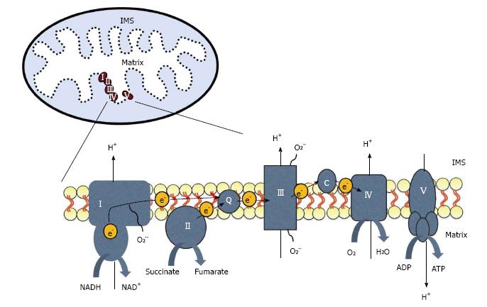
Figure 2 The electron transport chain is located on the inner mitochondrial membrane.
Electrons (e-) enter through complex I or II and are transferred to complex III by coenzyme Q (Q). Electrons then move between complex III and IV by cytochrome c (c). Electrons can leak from the chain and react with oxygen to create superoxide (O2-). The movement of electrons is coupled to the pumping of protons (H+) from the mitochondrial matrix into the intermembrane space (IMS). This gradient is then used by ATP synthase, or complex V, to generate ATP.
- Citation: O’Brien LC, Gorgey AS. Skeletal muscle mitochondrial health and spinal cord injury. World J Orthop 2016; 7(10): 628-637
- URL: https://www.wjgnet.com/2218-5836/full/v7/i10/628.htm
- DOI: https://dx.doi.org/10.5312/wjo.v7.i10.628


