Copyright
©The Author(s) 2016.
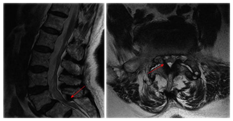
Figure 1 Magnetic resonance imaging of the spine found central lumbar spinal stenosis at L5/S1 level and L4-S1 foraminal narrowing.
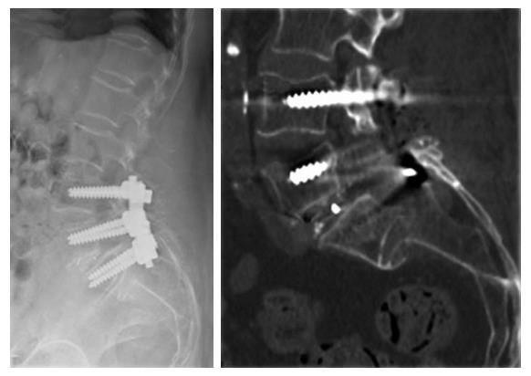
Figure 2 A computed tomography scan revealed a horizontal fracture at the S1/S2 level with S2 being totally displaced.
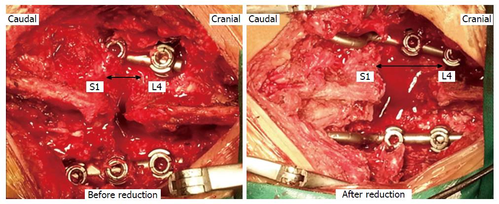
Figure 3 The interspinous process of the sacrum was totally displaced.
It moved backward and cranially onto the back of the instruments. To reduce the fracture, we used a spreader to distract the spinal processes of S1 and L4, which was effective. After the distraction, the distance between the spinal processes of the S1 and L4 increased significantly.
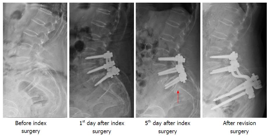
Figure 4 Sacral fracture found on the 5th day after surgery.
Two weeks after the index operation, the fusion construct was extended to the iliac wings using iliac screws.
- Citation: Wang Y, Liu XY, Li CD, Yi XD, Yu ZR. Surgical treatment of sacral fractures following lumbosacral arthrodesis: Case report and literature review. World J Orthop 2016; 7(1): 69-73
- URL: https://www.wjgnet.com/2218-5836/full/v7/i1/69.htm
- DOI: https://dx.doi.org/10.5312/wjo.v7.i1.69


