Copyright
©The Author(s) 2016.
Figure 1 Patient positioning.
A: Use of a regular table with bump under the sacrum and ability to lower the distal end of the bed down to afford better femoral exposure. An extra arm board can be placed on the contralateral distal end of the bed to support the contralateral leg while accessing the operative femoral canal; B: Patient positioning on a fracture-type table (Hana table, Mizuho Orthopedics Systems, Inc.).
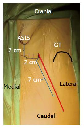
Figure 2 Surface anatomy for the direct anterior approach.
A 6-8 cm oblique incision is typically used by the authors. This incision may be extended proximally and distally as needed along the Smith-Petersen interval for adequate femoral and acetabular exposure.
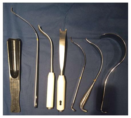
Figure 3 Selected retractors used for direct anterior approach.
From left to right, hip skid for ceramic head reduction, greater trochanteric retractor/elevator, femoral elevator for medial/calcar exposure (front and side views), medial acetabular wall retractor, posterior acetabular wall retractor, and tensor fascia lata retractor.
- Citation: Connolly KP, Kamath AF. Direct anterior total hip arthroplasty: Literature review of variations in surgical technique. World J Orthop 2016; 7(1): 38-43
- URL: https://www.wjgnet.com/2218-5836/full/v7/i1/38.htm
- DOI: https://dx.doi.org/10.5312/wjo.v7.i1.38


