Copyright
©The Author(s) 2015.
World J Orthop. Oct 18, 2015; 6(9): 744-749
Published online Oct 18, 2015. doi: 10.5312/wjo.v6.i9.744
Published online Oct 18, 2015. doi: 10.5312/wjo.v6.i9.744
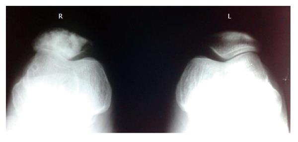
Figure 1 Radiograph of the right patella shows multiple lytic and sclerotic lesions involving the anterior two-thirds of the patella.
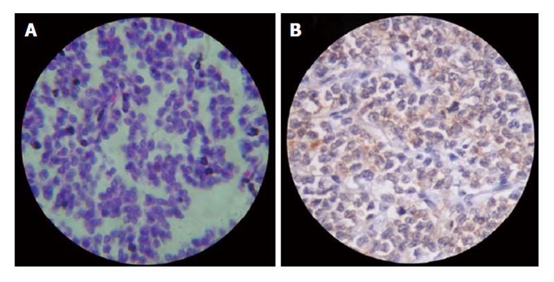
Figure 2 Haematoxylinand Eosin staining shows uniform, small, polygonal cells with scanty cytoplasm and indistinct cell borders (HE × 400) and Immunohistochemical staining shows CD99 diffuse positivity (A and B).
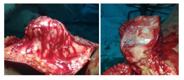
Figure 3 Peroperative picture shows thickened patella with uninvolved articular surface.
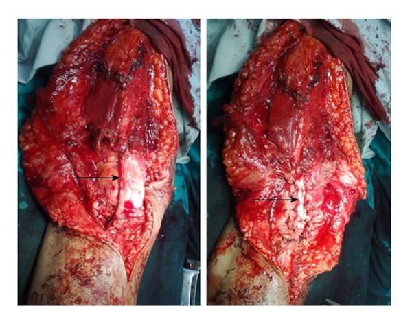
Figure 4 Peroperative picture shows repair of the extensor mechanism using V-Y plasty and split tendoachillesautograft.
- Citation: Valsalan RM, Zacharia B. Ewings sarcoma of patella: A rare entity treated with a novel technique of extensor mechanism reconstruction using tendoachilles auto graft. World J Orthop 2015; 6(9): 744-749
- URL: https://www.wjgnet.com/2218-5836/full/v6/i9/744.htm
- DOI: https://dx.doi.org/10.5312/wjo.v6.i9.744


