Copyright
©The Author(s) 2015.
World J Orthop. Sep 18, 2015; 6(8): 641-648
Published online Sep 18, 2015. doi: 10.5312/wjo.v6.i8.641
Published online Sep 18, 2015. doi: 10.5312/wjo.v6.i8.641
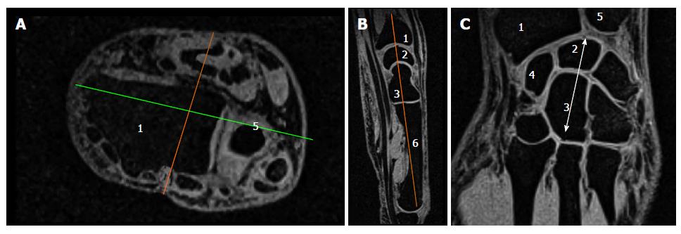
Figure 1 Standardized selection of a slice of interest.
A: Axial slice of the wrist in which we initially determined the axis of the anterior part of the Radius (1) (green line) and then the perpendicular sagittal axis (orange line); B: Corresponding sagittal slice in which, we chose the slice going through the proximal part of the capitate (3) and the metacarpal basis (6); C: Corresponding coronal slice showing the radius (1), ulna (5), navicular (4), semi-lunar (2) and capitate bones (3). Arrow indicates the carpal bone length measurement from the lowest point of the capitate to the highest point of the semi-lunar bone.
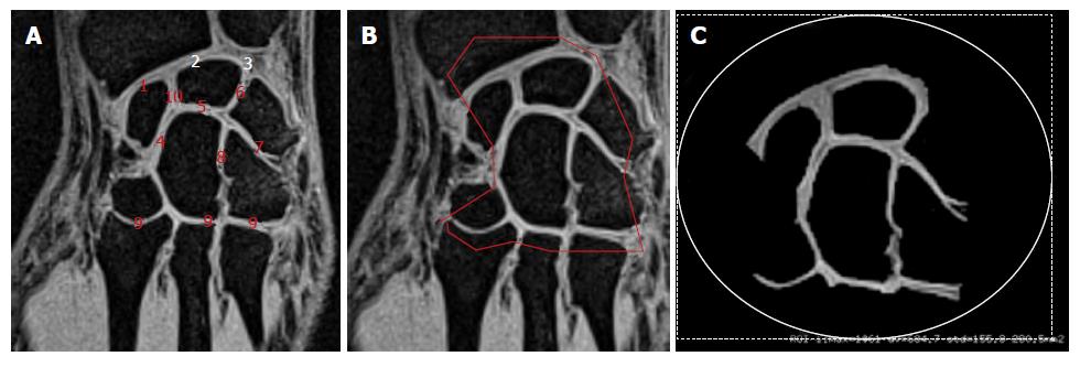
Figure 2 Manual segmentation of the cartilage cross-sectional area.
A: Coronal slice (as in Figure 1C) showing different joints between radius and scaphoid (1), radius and lunate (2), lunate and ulna (3), scaphoid and capitate (4), lunate and ulna (5), lunate and triquetrum (6), triquetrum and hamate (7) hamate and capitate (8), carpals bones and metacarpal bones (9) scaphoid and lunate (10); B: Coronal slice illustrating the manual segmentation of the cartilage area of interest and the corresponding result (C).
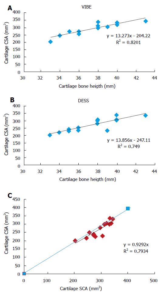
Figure 3 Relationships between wrist cartilage cross-sectional area measurements.
Relationship between wrist cartilage CSA (mm2) and carpal bone length (mm) measured. The cartilage CSA was measured using the VIBE (A) and the DESS (B) MRI sequence; C: Relationship between cartilage CSA measured using the VIBE (x-axis) and the DESS (y-axis) MRI sequences. The continuous line crossing the both axes represents the identity line. VIBE: Volume interpolated breath hold examination; DESS: Dual echo steady state; CSA: Cartilage cross-sectional area; MRI: Magnetic resonance imaging.
- Citation: Zink JV, Souteyrand P, Guis S, Chagnaud C, Fur YL, Militianu D, Mattei JP, Rozenbaum M, Rosner I, Guye M, Bernard M, Bendahan D. Standardized quantitative measurements of wrist cartilage in healthy humans using 3T magnetic resonance imaging. World J Orthop 2015; 6(8): 641-648
- URL: https://www.wjgnet.com/2218-5836/full/v6/i8/641.htm
- DOI: https://dx.doi.org/10.5312/wjo.v6.i8.641


