Copyright
©The Author(s) 2015.
World J Orthop. Sep 18, 2015; 6(8): 590-601
Published online Sep 18, 2015. doi: 10.5312/wjo.v6.i8.590
Published online Sep 18, 2015. doi: 10.5312/wjo.v6.i8.590
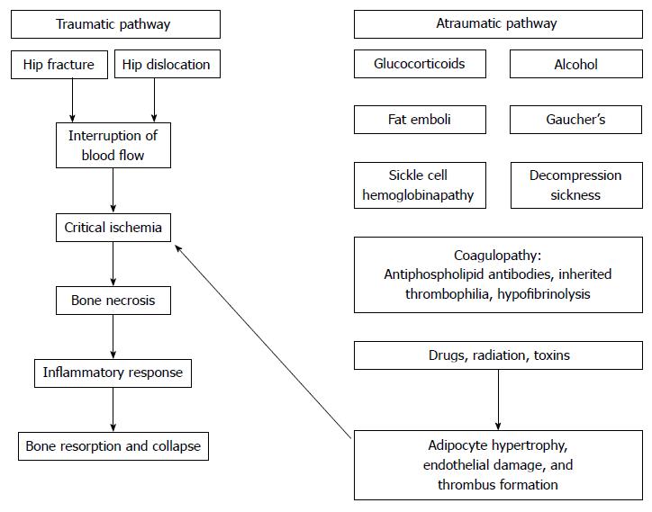
Figure 1 Mechanisms of osteonecrosis.
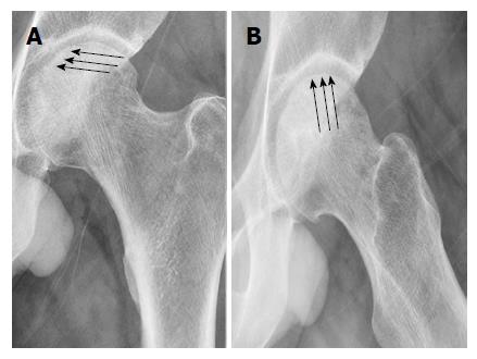
Figure 2 Left hip anteriorposterior and cross leg lateral X-rays showing (arrows) the crescent sing.
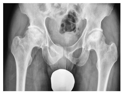
Figure 3 Bilateral osteonecrosis of the femoral head with flattening of the surface and early sings of osteoarthritis.
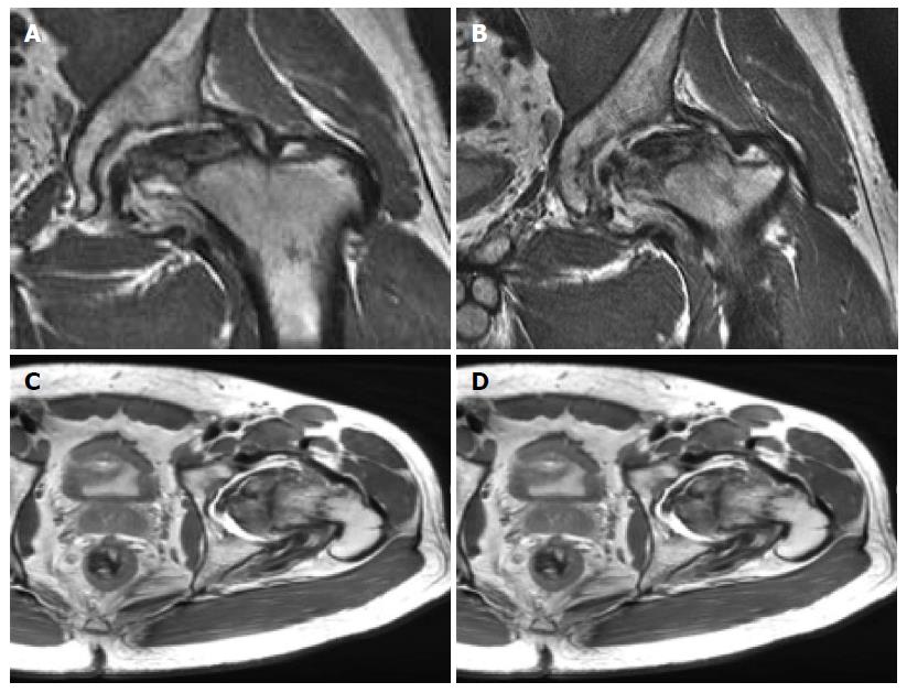
Figure 4 Magnetic resonance imaging of the left hip showing extensive avascular necrosis of the femoral head with collapse and a large area of devitalized bone demonstrating fibrocystic change.
There is associated severe arthrosis of the left hip joint with a moderate effusion, synovitis and debris and a marked bone marrow edema pattern on both sides of the joint.
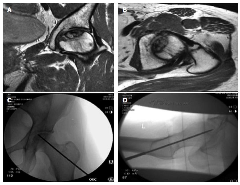
Figure 5 Core decompression of the left femoral head.
Preoperative magnetic resonance imaging, above (coronal and axial views) and fluoroscopic imaging during the procedure below.
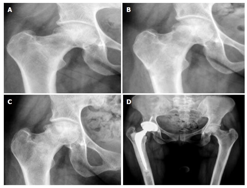
Figure 6 Right femoral head osteonecrosis.
Flattening of femoral head progression in 24 mo ending up in a right total hip replacement.
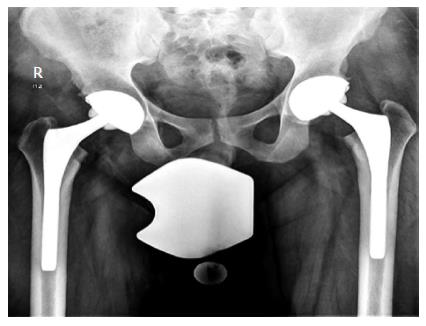
Figure 7 Bilateral total hip replacement in a patient with bilateral hip osteonecrosis of the femoral head.
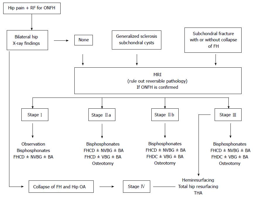
Figure 8 Algorithm for the management and treatment of patients with osteonecrosis of the femoral head.
RF: Risk factors; ONFH: Osteonecrosis of the femoral head; FH: Femoral head; MRI: Magnetic resonance imaging; FHCD: Femoral head core decompression; NVBG: Non vascularized bone graft; BA: Biologic agents; VBG: Vascularized bone graft; OA: Osteoarthritis; THA: Total hip arthroplasty.
- Citation: Moya-Angeler J, Gianakos AL, Villa JC, Ni A, Lane JM. Current concepts on osteonecrosis of the femoral head. World J Orthop 2015; 6(8): 590-601
- URL: https://www.wjgnet.com/2218-5836/full/v6/i8/590.htm
- DOI: https://dx.doi.org/10.5312/wjo.v6.i8.590


