Copyright
©The Author(s) 2015.
World J Orthop. Dec 18, 2015; 6(11): 886-901
Published online Dec 18, 2015. doi: 10.5312/wjo.v6.i11.886
Published online Dec 18, 2015. doi: 10.5312/wjo.v6.i11.886
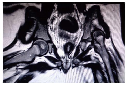
Figure 1 Magnetic resonance imaging of a two-year-old girl with developmental dysplasia of the hip.
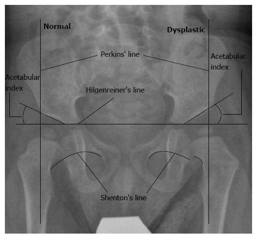
Figure 2 One-year-old girl with developmental dysplasia of the hip on the left side.
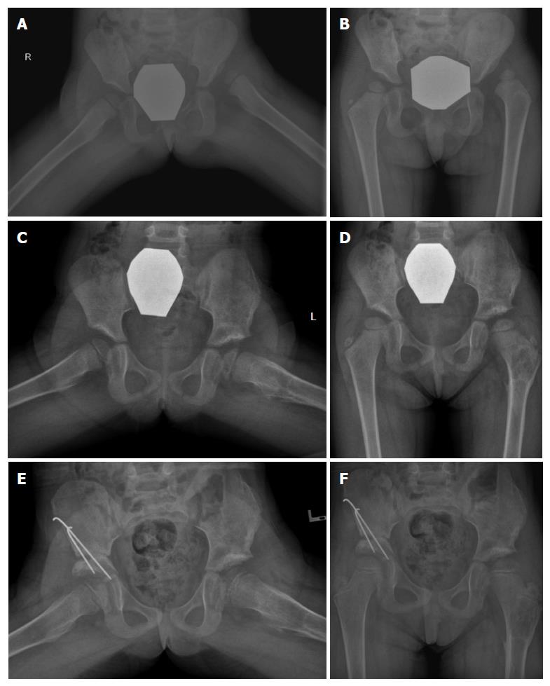
Figure 3 Hip reconstruction.
A, B: Female patient, diagnosed with bilateral developmental dysplasia of the hip at the age of 2.5 years; C, D: On presentation left hip reconstruction, that included open reduction, Salter osteotomy and femoral derotational osteotomy with shortening, was performed. Post-operative X-rays after removal of the internal fixation devices are shown; E, F: Due to persistent mild hip dysplasia on the right side, a Salter osteotomy was performed at the age of 4 years.
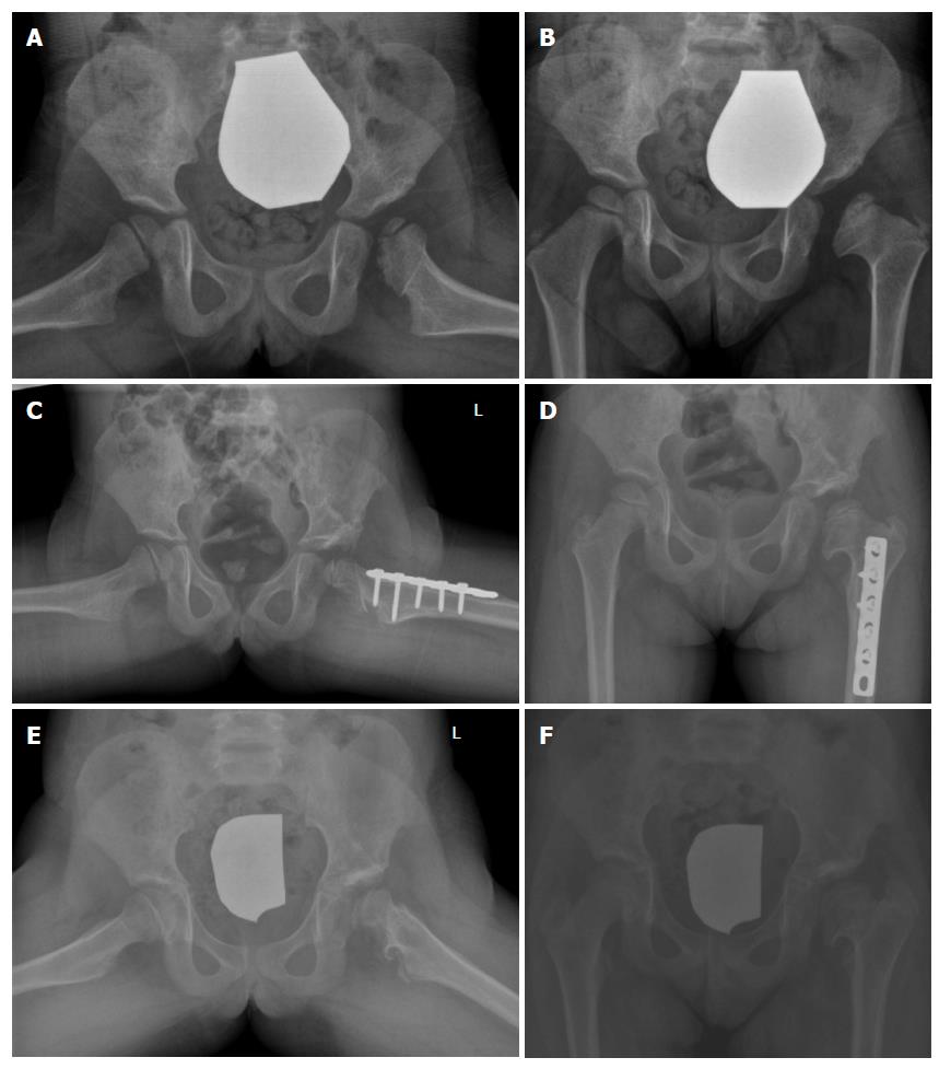
Figure 4 Acetabuloplasty.
A female patient was diagnosed with DDH of the left hip at the age of 6 mo using sonography. She was treated with a spica cast. AVN of the left femoral head was seen on follow-up radiographs at the age of 3 years (A,B); C, D: Acetabuloplasty of the left hip with femoral derotational osteotomy was performed at the age of 5 years; E, F: Follow-up X-rays at the age of 8 years. DDH: Developmental dysplasia of the hip; AVN: Avascular necrosis.
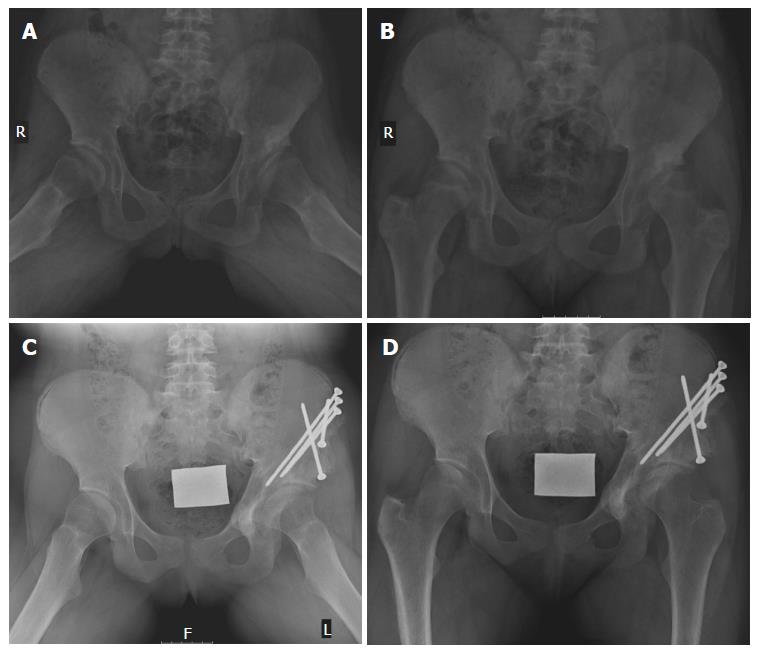
Figure 5 Female patient with developmental dysplasia of the hip that was diagnosed in early adolescence.
A, B: X-rays at the time of diagnosis of DDH at the age of 13 years; C, D: X-rays taken two years after Ganz osteotomy on the left side. DDH: Developmental dysplasia of the hip.
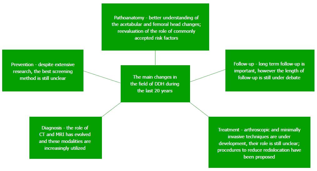
Figure 6 Summary of the main changes in the field of developmental dysplasia of the hip during the last 20 years.
MRI: Magnetic resonance imaging; DDH: Developmental dysplasia of the hip; CT: Computed tomography.
- Citation: Kotlarsky P, Haber R, Bialik V, Eidelman M. Developmental dysplasia of the hip: What has changed in the last 20 years? World J Orthop 2015; 6(11): 886-901
- URL: https://www.wjgnet.com/2218-5836/full/v6/i11/886.htm
- DOI: https://dx.doi.org/10.5312/wjo.v6.i11.886


