Copyright
©The Author(s) 2015.
World J Orthop. Nov 18, 2015; 6(10): 829-837
Published online Nov 18, 2015. doi: 10.5312/wjo.v6.i10.829
Published online Nov 18, 2015. doi: 10.5312/wjo.v6.i10.829
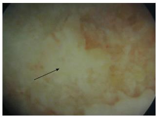
Figure 1 Endoscopic view of the osteonecrotic lesion.
View obtained from the endoscopy through the canal. The posterior aspect of the osteonecrotic lesion is seen at the center of the image (arrow) as a white non-vascularized area, surrounded by normal purple-coloured bone (vascularized bone).
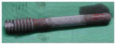
Figure 2 Tantalum rod.
The tantalum rod used in the present series, manufactured by Zimmer (Zimmer, Inc, Warsaw, IN, United States). The rod was impregnated with bone marrow aspirates obtained from iliac crest aspiration.
Figure 3 Pre- and post-operative imaging of femoral head osteonecrosis.
Imaging of a 42-year-old patient with corticosteroid-induced osteonecrosis of the femoral head treated with porous tantalum. A: Preoperative MRI of Steinberg stage II osteonecrosis of the left femoral head; B: One-year postoperative X-ray after the implantation of the tantalum rod. No pathological findings are seen; C: Five-year postoperative MRI of the left hip with absence of pathological findings. MRI: Magnetic resonance imaging.
Figure 4 Pre- and postoperative X-rays of osteonecrosis of the femoral head.
X-rays of a 47-year-old patient with idiopathic osteonecrosis of the femoral head. A: Preoperative X-ray with Steinberg stage II osteonecrosis; B: Six-month postoperative X-ray showing no disease deterioration; C: Five-year postoperative X-ray of the same patient with no pathological findings.
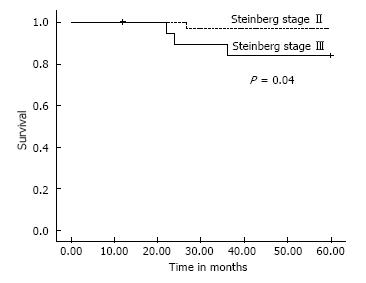
Figure 5 Survival plots for patients with stage II and stage III disease.
Within the 5-year follow up period, 4 patients with osteonecrosis treated with tantalum rod were converted to total hip arthroplasty. One patient had Steinberg stage II disease and 3 patients had Steinberg stage III disease. Patients with stage III disease had increased risk to undergo total hip arthroplasty. The difference was statistically significant (P = 0.04).
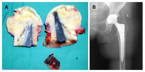
Figure 6 Conversion of tantalum rod to total hip arthroplasty.
A: The osteotomised femoral head with the tantalum rod in a 55-year-old patient who underwent THA due to disease deterioration. The osteotomy of the femoral neck was performed through the tantalum rod; B: Six-week post-THA X-ray of the same patient. The X-ray shows callous formation within the trochanteric hole. THA: Total hip arthroplasty.
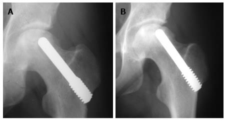
Figure 7 Disease progression after tantalum rod implantation.
Thirty-eight-year old female patient with osteonecrosis of the left femoral head due to leukemia. A: The immediate postoperative X-ray after the implantation of tantalum rod showing Steinberg stage III osteonecrosis; B: X-ray of the same patient showing deterioration to Steinberg stage IV osteonecrosis 12 mo after the implantation of tantalum rod.
- Citation: Pakos EE, Megas P, Paschos NK, Syggelos SA, Kouzelis A, Georgiadis G, Xenakis TA. Modified porous tantalum rod technique for the treatment of femoral head osteonecrosis. World J Orthop 2015; 6(10): 829-837
- URL: https://www.wjgnet.com/2218-5836/full/v6/i10/829.htm
- DOI: https://dx.doi.org/10.5312/wjo.v6.i10.829


