Copyright
©The Author(s) 2015.
World J Orthop. Nov 18, 2015; 6(10): 804-811
Published online Nov 18, 2015. doi: 10.5312/wjo.v6.i10.804
Published online Nov 18, 2015. doi: 10.5312/wjo.v6.i10.804
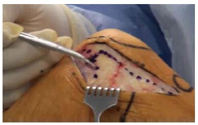
Figure 1 The incisions for minimally invasive surgery of the knee.
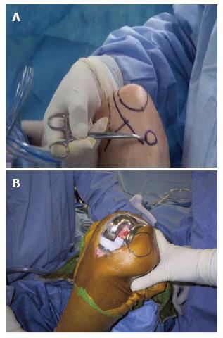
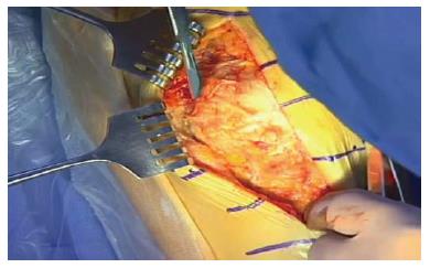
Figure 2 Anatomic location of the incision.
A: The skin outline of the minimally invasive surgery incision and its relationship to the patella, the medial femoral condyle, and the tibial joint line; B: Total knee arthroplasty with the quadriceps sparing approach.
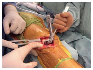
Figure 3 Specially designed instruments for minimally invasive surgery total knee arthroplasty.
This instrument resects the anterior aspect of the femur in full extension.
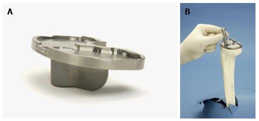
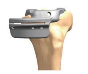
Figure 4 Implants designed specifically for minimally invasive surgery total knee arthroplasty.
A: A limited keel on a modified tibial tray for MIS; B: A dropdown stem used with a modular tibial tray.
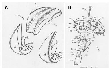
Figure 5 Modular components for minimally invasive surgery total knee arthroplasty.
A: Modular femoral component; B: Modular tibial component.
Figure 6 Mini-midvastus arthrotomy.
The dotted line shows the medial arthrotomy with the continuation of the incision into the vastus medialis muscle splitting the distal one third of the muscle from the proximal two thirds in the line of the fibers.
Figure 7 Subvastus arthrotomy.
The subvastus incision along the inferior border of the vastus medialis muscle. The scalpel blade is along the inferior edge of the vastus medialis and will extend the arthrotomy from there distally along the medial side of the patella and the patellar tendon.
Figure 8 Cutting blocks for patient specific instrumentation.
A patient specific cutting block for tibial resection.
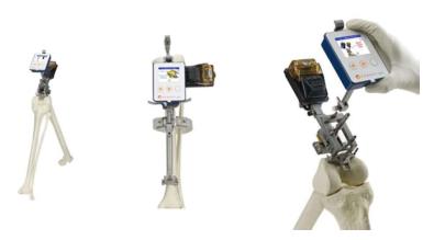
Figure 9 Smart instruments for alignment of total knee arthroplasty cuts.
Orthalign™ (Orthalign, Aliso Viejo, California, United States) instrument for femoral and tibial resection.
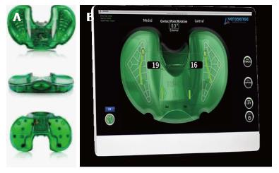
Figure 10 Sensor devices for total knee arthroplasty.
A: Orthosensor™ (Verasense, Orthosensor, Dania Beach, Florida, United States) inserts for the modular trial tibia; B: Pressure recording from the Orthosensor™ plate.
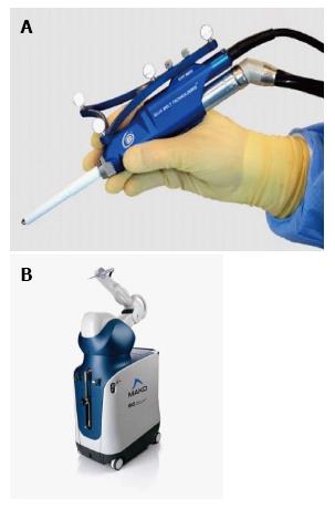
Figure 11 Haptic instrumentation for total knee arthroplasty.
A: Navio™ (Blue Belt Technologies, Plymouth, MN, United States) robotic hand piece that is a haptic control. The surgeon moves the cutting instrument and the computer turns the device off if the resection area is exceeded or the depth is too great; B: MAKOTM (MAKO surgical corporation, Fort Lauderdale, Florida, United States) robotic arm. A haptic arm that is moved by the surgeon within the confines of the resection area and within the proper depth.
- Citation: Tria AJ, Scuderi GR. Minimally invasive knee arthroplasty: An overview. World J Orthop 2015; 6(10): 804-811
- URL: https://www.wjgnet.com/2218-5836/full/v6/i10/804.htm
- DOI: https://dx.doi.org/10.5312/wjo.v6.i10.804


