Copyright
©2014 Baishideng Publishing Group Inc.
World J Orthop. Nov 18, 2014; 5(5): 614-622
Published online Nov 18, 2014. doi: 10.5312/wjo.v5.i5.614
Published online Nov 18, 2014. doi: 10.5312/wjo.v5.i5.614
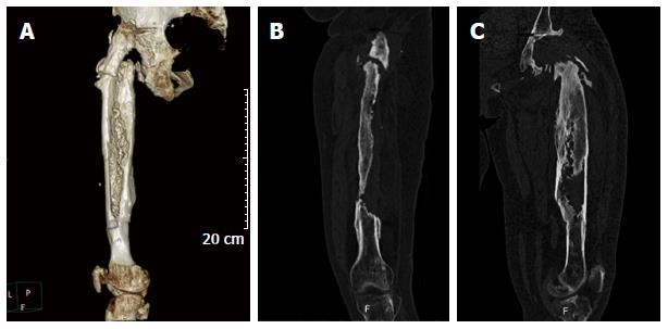
Figure 1 Computed tomography scan images with metallic artifact subtraction and three-dimensional reconstruction consist a useful tool for precise assessment of the amount of bone loss and the specific variations of the femoral anatomy preoperatively.
A: Computed tomography scan image of the left femur (posterior projection) with metallic artifact subtraction and three-dimensional reconstruction showing precisely the amount of bone loss of the proximal part of the femur; B: Coronal; C: Sagittal view of the same case.
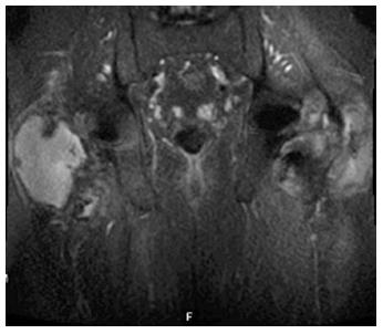
Figure 2 Metal artifact reduction magnetic resonance imaging (coronal view) showing the adverse reactions to metal debris and formation of pseudocapsules in both hips following bilateral total hip arthroplasties with modular necks.
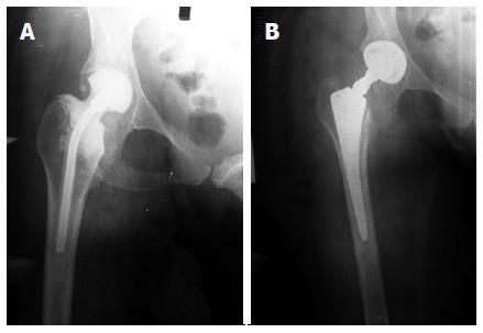
Figure 3 Anteroposterior radiograph of bone loss.
A: Anteroposterior (AP) radiograph of the right hip showing minimal bone loss of the proximal metaphyseal bone secondary to periprosthetic hip infection. A antibiotic cement spacer was implanted after irrigation and debridement during the first stage of a two-stage exchange arthroplasty; B: AP radiograph of the same hip after the second stage. The metaphyseal bone loss was minimal and a cementless primary stems with common length and geometry was used.
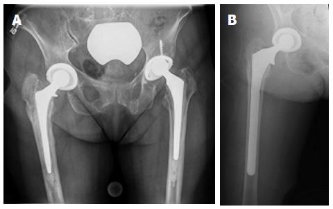
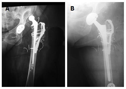
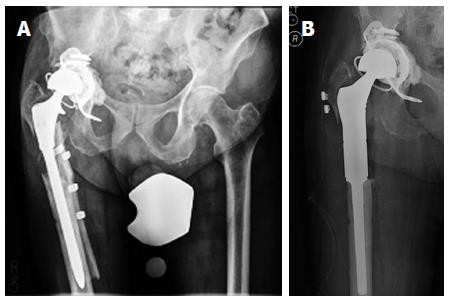
Figure 4 Extensively porous coated stems.
A: Anteroposterior (AP) radiograph of the pelvis demonstrating bilateral total hip arthroplasties. In right hip there is evident loosening of the femoral stem and extensive metaphyseal cancellous bone loss with some diaphyseal bone loss, which is limited to less than 4 cm of diaphyseal bone; B: AP radiograph of the same hip post revision using an extensively porous coated stem.
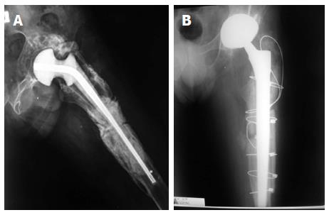
Figure 5 Modular stems.
A: Preoperative anteroposterior (AP) radiograph of a dislocated left hip with a paprosky type IIIA defect; B: Postoperative AP radiograph of the revised hip with a modular stem.
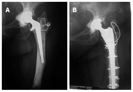
Figure 6 Newer stem designs with modular configuration have been associated with lower rates of subsidence and improved restoration of limb length and femoral offset.
A: Anteroposterior (AP) radiograph of the left hip showing extensive bone loss of the proximal metaphyseal bone with significant diaphyseal bone loss (Paparosky type IIIB) secondary to periprosthetic hip infection. A antibiotic cement spacer was implanted after irrigation and debridement during the first stage of a two-stage exchange arthroplasty; B: AP radiograph of the same hip after the second stage using a distal fixation taper fluted stem.
Figure 7 Multiple osteotomies allow for restoration of the anatomical axis of the femur, an easier access of the distal segment of the modular stem, thus reducing the risk of femoral fracture or perforation of the cortex.
A: Anteroposterior (AP) radiograph of the left hip showing a cemented femoral component that is failed in varus, resulting in a slight angular malalignment of femur; B: AP radiograph of the same hip after revision of the femoral component using a modular stem, which combines both proximal metaphyseal and distal fixation due to the taper design and the distal flutes. A healed corrective osteotomy at the level of the mid-diaphysis facilitated the insertion of the stem and correction of the angular deformity to its neutral axis.
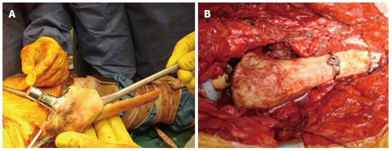
Figure 8 The allograft is reamed and broached and a long stem is cemented at the back table.
A: Intraoperative picture demonstrating the allograft-prosthesis composite preparation. An allograft of appropriate size is osteotomized at the desired subtrochanteric level in order to match the bony defect of the proximal femur. The allograft is reamed and broached and a long stem is cemented at the back table; B: Intraoperative picture showing the allograft-prosthesis composite with a lateral sleeve that offers a wide area of bone contact with the distal host femur. Circlage cables are used to secure the allograft-host bone fixation.
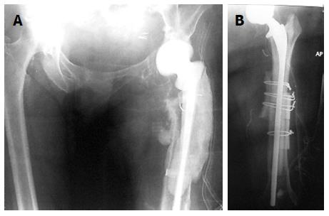
Figure 9 The allograft-prosthesis composite is implanted to the native femur with the use of cement or not, depending on the selected type of implant and the quality of host bone.
A: Anteroposterior (AP) radiograph of the pelvis demonstrating a Paprosky type IV femoral defect of the left hip as a result of a periprosthetic infection. The femoral canal is widened and there is no sufficient diaphyseal support for future cementless fixation; B: Postoperative AP radiograph of the left hip showing reconstruction of the proximal femoral defect with the use of an allograft-prosthesis composite. Remnants of the host bone are wrapped-around the femur at the level of the allograft-host bone junction in order to improve incorporation of the allograft to the host femur.
Figure 10 Proximal femur replacement using the so-called “mega-prostheses” is an alternative option in cases of severe proximal femoral bone loss.
A: Anteroposterior (AP) radiograph of the pelvis showing a left revision total hip arthroplasty in a 82-year-old male patient with an extensive structural defect of the lateral femoral cortex resulting in a painful and mechanically loose construct of a periprosthetic hip fracture; B: Postoperative AP radiograph of the right hip showing the replacement of the proximal femoral with a megaprosthesis.
- Citation: Sakellariou VI, Babis GC. Management bone loss of the proximal femur in revision hip arthroplasty: Update on reconstructive options. World J Orthop 2014; 5(5): 614-622
- URL: https://www.wjgnet.com/2218-5836/full/v5/i5/614.htm
- DOI: https://dx.doi.org/10.5312/wjo.v5.i5.614


