Copyright
©2014 Baishideng Publishing Group Inc.
World J Orthop. Jul 18, 2014; 5(3): 292-303
Published online Jul 18, 2014. doi: 10.5312/wjo.v5.i3.292
Published online Jul 18, 2014. doi: 10.5312/wjo.v5.i3.292
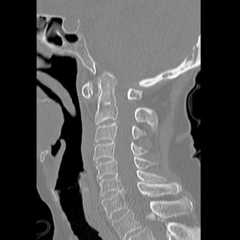
Figure 1 Sagittal computed tomography of the cervical spine of an 82-year-old female with rheumatoid arthritis and neck pain with cranial settling.
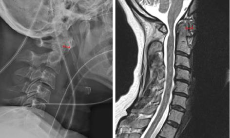
Figure 2 Lateral X-ray (left) and sagittal magnetic resonance imaging (right) of a 52-year-old with atlantoaxial subluxation.
Left: Lateral X-ray; Right: Sagittal Magnetic resonance imaging. The anterior atlantodental interval is shown (red line).
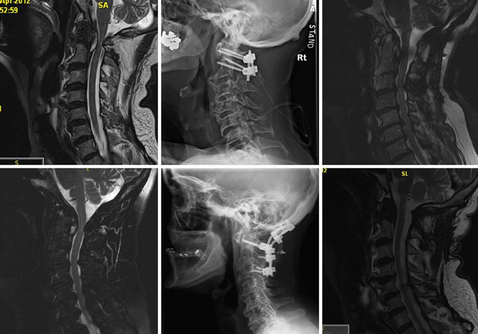
Figure 3 Resolving rheumatoid pannus after occipital cervical (top) and C1-2 fusion (bottom).
Left: Preoperative magnetic resonance imagings; Middle: Postoperative lateral X-ray; Right: Postoperative magnetic resonance imagings.
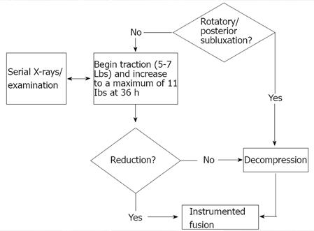
Figure 4 Preoperative traction and surgical approach to cervical rheumatoid.
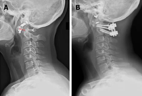
Figure 5 Lateral X-ray images of the spine before (A) and after (B) surgical intervention.
The anterior atlantodental interval is shown (red line).
- Citation: Mallory GW, Halasz SR, Clarke MJ. Advances in the treatment of cervical rheumatoid: Less surgery and less morbidity. World J Orthop 2014; 5(3): 292-303
- URL: https://www.wjgnet.com/2218-5836/full/v5/i3/292.htm
- DOI: https://dx.doi.org/10.5312/wjo.v5.i3.292


