Copyright
©2014 Baishideng Publishing Group Co.
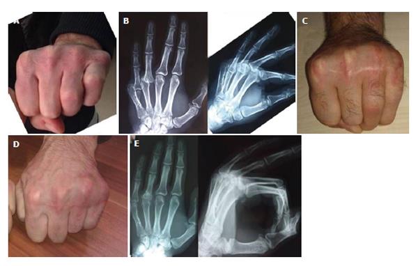
Figure 1 Preoperative and postoperative clinical radiological images of case 1.
A: Pre-operative 30° limitation of flexion in the 4th and 60° limitation of flexion in the 5th metacarpophalangeal (MP) joint; B: Preoperative AP and lateral radiographies; C: 4th MP joint has 20° and 5th MP joint has 30° limitation of flexion at 2 mo postoperative; D: Full flexion at the 4th MP joint and 10 degrees limitation of flexion at the 5th MP joint at postoperative 12th month; E: Radiographies at 12th month. The reduction is maintained and the osteotomy line at the 4th proximal phalanx had fused.
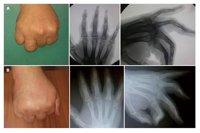
Figure 2 Preoperative and postoperative clinical radiological images of case 2.
A: Preoperative 70 degrees of limitation of flexion at the 4th metacarpophalangeal joint. There is a deformity dorsal to the proximal phalanx proximal joint surface; B: 30 degrees of limitation of flexion and union of the proximal phalangeal osteotomy line at 8th month control.
Figure 3 Exposure of the dislocated metacarpophalangeal joint with a dorsal approach.
Because of the deformation of the dorsal joint surface of the 4th proximal phalanx, a volar closing wedge osteotomy was done, which was then stabilized with a K wire.
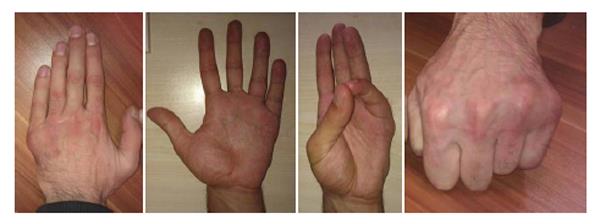
Figure 4 Full flexion at the 4th metacarpophalangeal joint and 10 degrees limitation of flexion at the 5th metacarpophalangeal joint at postoperative 12th month.
The 4th and 5th metacarpophalangeal joints have full extension.
- Citation: Başar H, İnanmaz ME, Köse K&, Tetik C. Isolated dorsal approach for the treatment of neglected volar metacarpophalangeal joint dislocations. World J Orthop 2014; 5(1): 62-66
- URL: https://www.wjgnet.com/2218-5836/full/v5/i1/62.htm
- DOI: https://dx.doi.org/10.5312/wjo.v5.i1.62


