Copyright
©2013 Baishideng Publishing Group Co.
World J Orthop. Oct 18, 2013; 4(4): 323-326
Published online Oct 18, 2013. doi: 10.5312/wjo.v4.i4.323
Published online Oct 18, 2013. doi: 10.5312/wjo.v4.i4.323
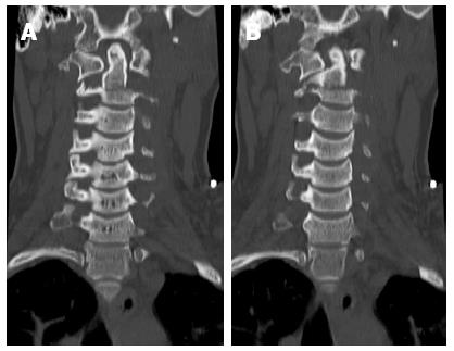
Figure 1 Reformatted coronal computer tomography images showing the transverse body fracture extending through the transverse processes (A, B).
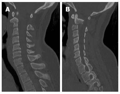
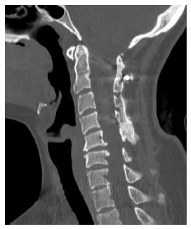
Figure 2 Reformatted sagittal computer tomography images right of midline (A), and left of midline (B).
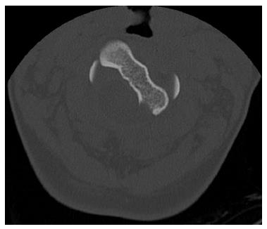
Figure 3 Axial computer tomography showing left rotational component of upper C2 body.
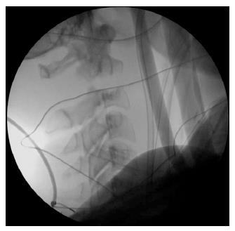
Figure 4 Lateral X-ray image showing persistent distraction in halo brace.
Figure 5 Reformatted sagittal computer tomography image obtained at 2-year follow-up showing anatomic alignment and fused segments.
- Citation: Lau D, Shin SS, Patel R, Park P. Treatment of C2 body fracture with unusual distractive and rotational components resulting in gross instability. World J Orthop 2013; 4(4): 323-326
- URL: https://www.wjgnet.com/2218-5836/full/v4/i4/323.htm
- DOI: https://dx.doi.org/10.5312/wjo.v4.i4.323


