Copyright
©2013 Baishideng Publishing Group Co.
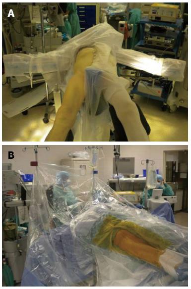
Figure 1 Patient positioned supine on a specialized orthopedic table (A) with the operative leg prepped and draped (B).
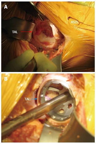
Figure 2 Acetabular exposure.
A: Acetabular exposure with the direct anterior approach; B: Acetabular exposure using the direct anterior approach with trial component in place. The transverse acetabular ligament (TAL) is clearly identified by arrow. ASIS: Anterior superior iliac spine.
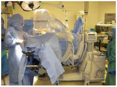
Figure 3 The supine position of the patient on the operating table facilitates the use fluoroscopy during surgery to assess component position and alignment.
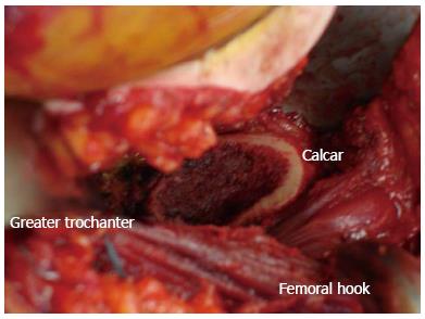
Figure 4 Exposure following femoral neck osteotomy with the direct anterior approach using a specialized orthopedic table.
- Citation: Moskal JT, Capps SG, Scanelli JA. Anterior muscle sparing approach for total hip arthroplasty. World J Orthop 2013; 4(1): 12-18
- URL: https://www.wjgnet.com/2218-5836/full/v4/i1/12.htm
- DOI: https://dx.doi.org/10.5312/wjo.v4.i1.12


