Copyright
©2012 Baishideng Publishing Group Co.
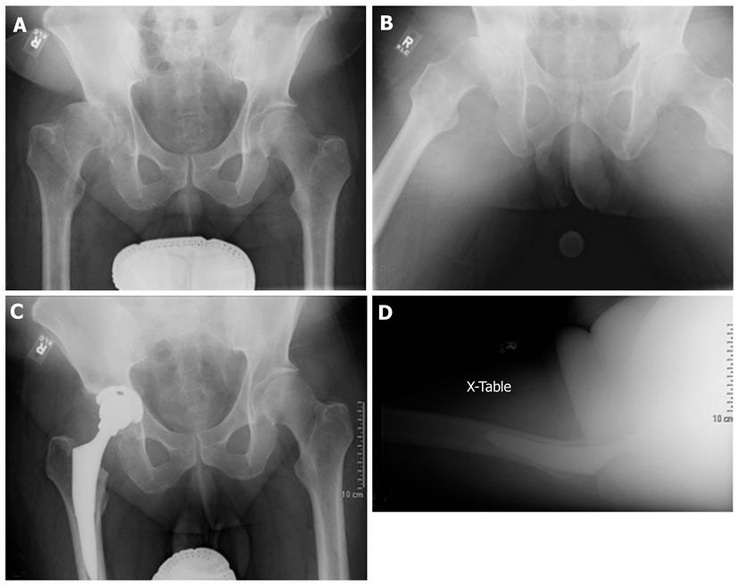
Figure 1 Treatment of Crowe I hip using an anatomic hip center.
A and B: Pre-op X-rays; C and D: Post-op X-rays.
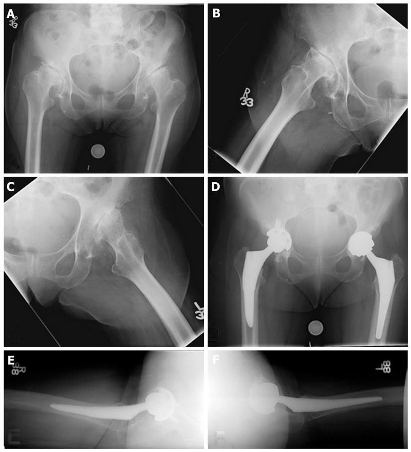
Figure 2 Treatment of Crowe II (right) and III (left) hips using an anatomic hip center with medialization.
A, B and C: Pre-op X-rays; D, E and F: Post-op X-rays.
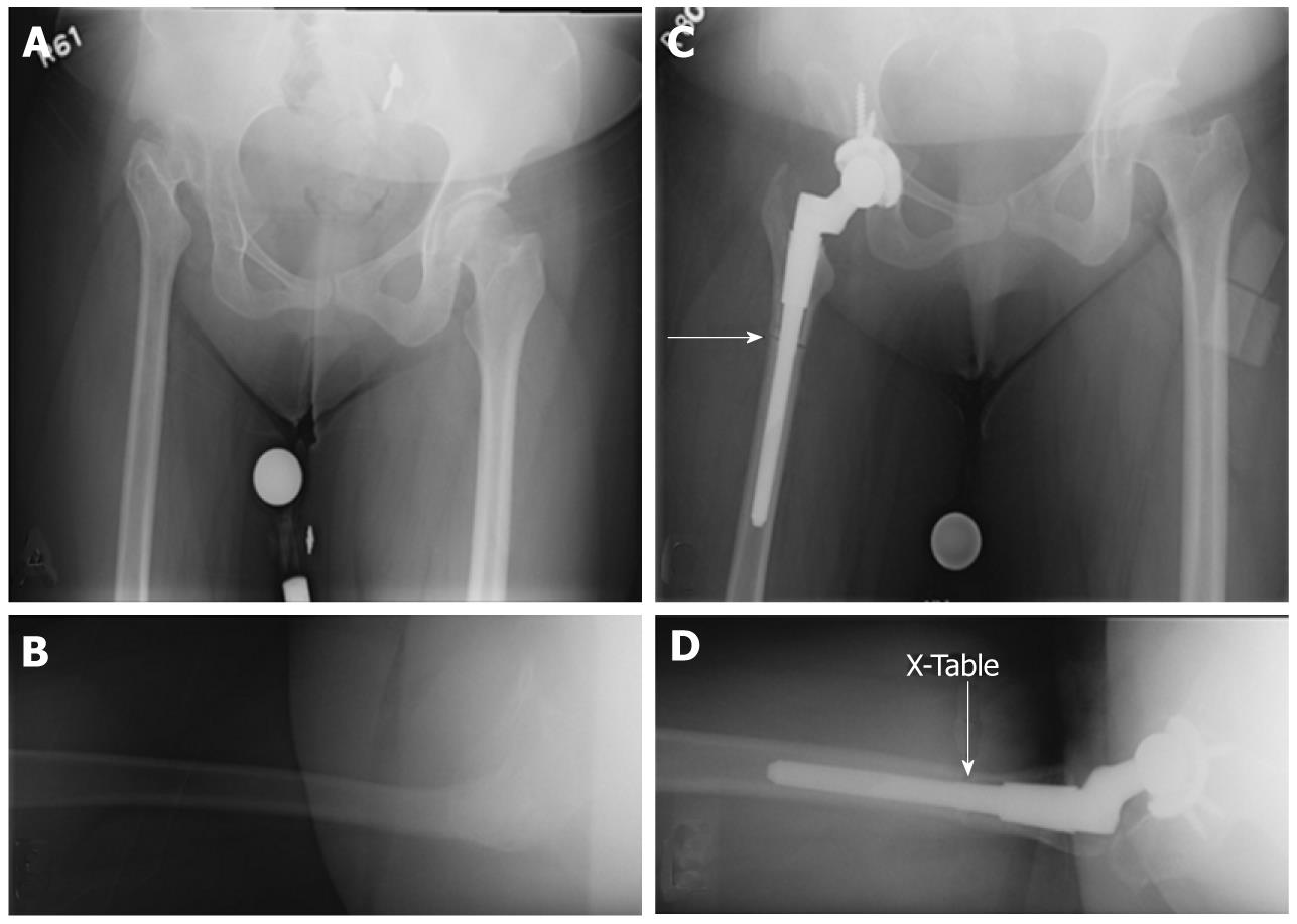
Figure 3 Treatment of Crowe IV hip using an anatomic hip center with subtrochanteric shortening osteotomy (arrow) and a modular femoral component.
A and B: Pre-op X-rays; C and D: Post-op X-rays.
- Citation: Yang S, Cui Q. Total hip arthroplasty in developmental dysplasia of the hip: Review of anatomy, techniques and outcomes. World J Orthop 2012; 3(5): 42-48
- URL: https://www.wjgnet.com/2218-5836/full/v3/i5/42.htm
- DOI: https://dx.doi.org/10.5312/wjo.v3.i5.42


