
















Copyright: ©Author(s) 2026.
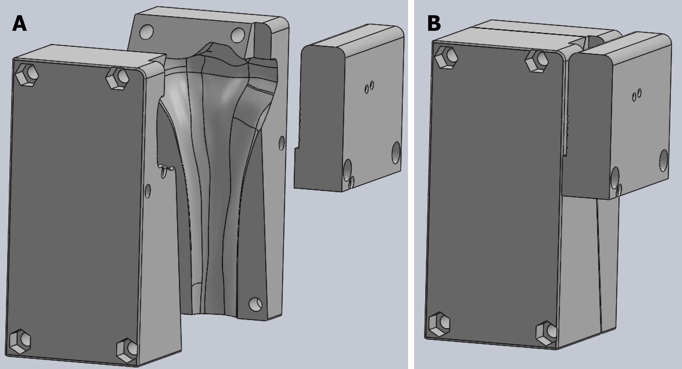
Figure 1 The 3D printed guides use for the reproducible generation of tibia plateau fractures.
A: Left; B: Closed.
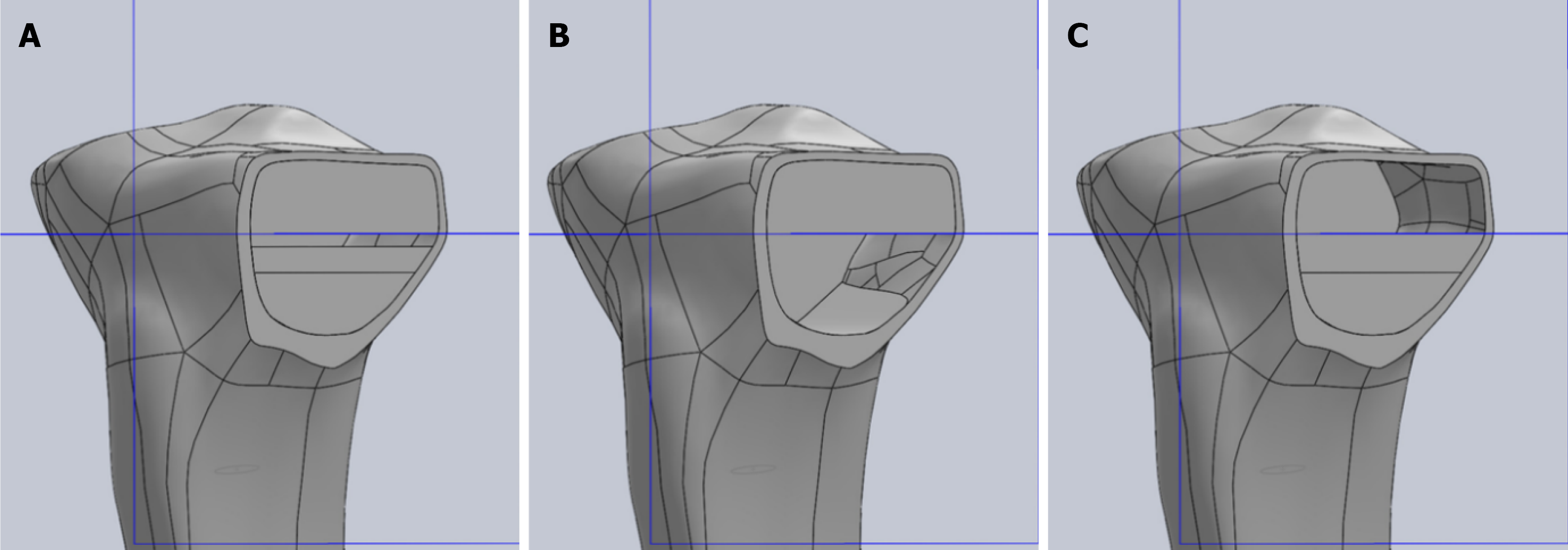
Figure 2 The three scenarios of bone deficit tested.
A: 10% distal deficit; B: 50% distal deficit; C: 50% proximal deficit.
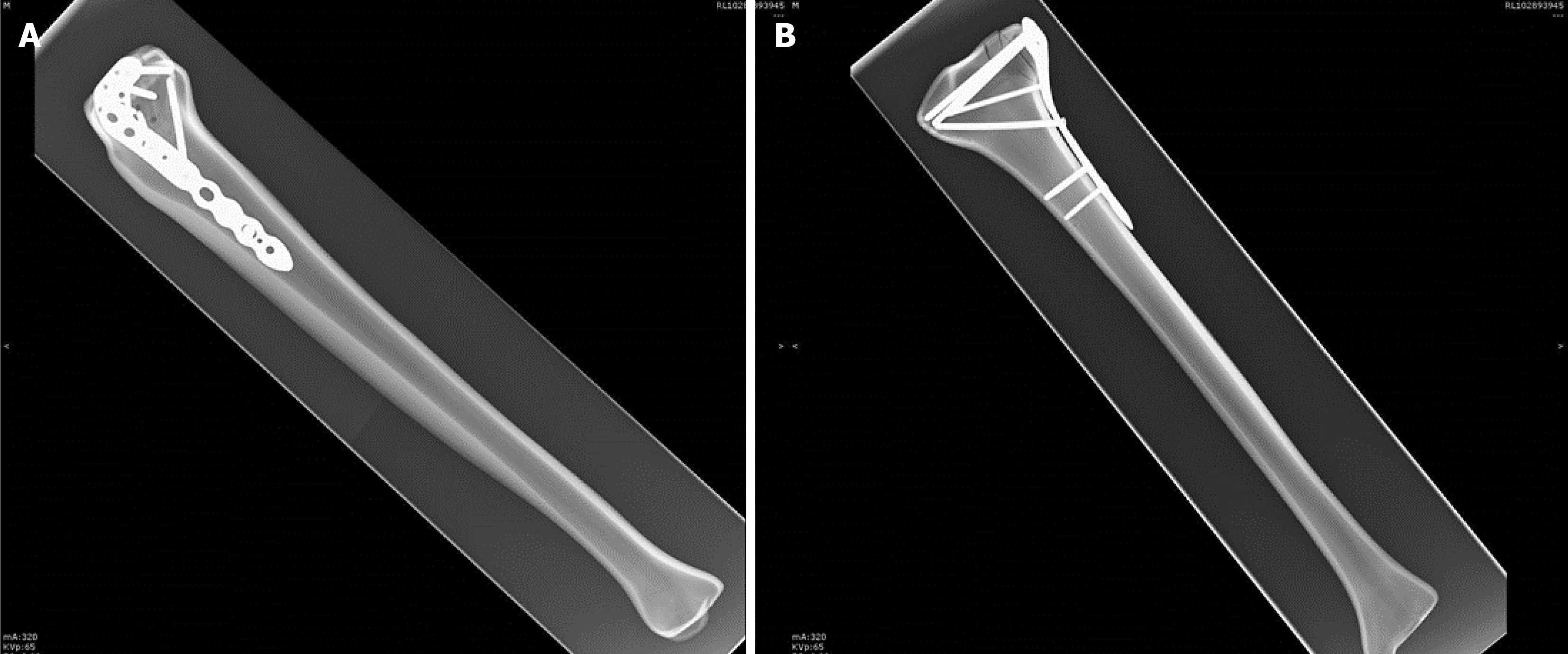
Figure 3 Saw bone Lateral tibia plateau fracture fixed with locking plate.
A: Lateral X-ray view; B: Anterior-posterior X-ray view.
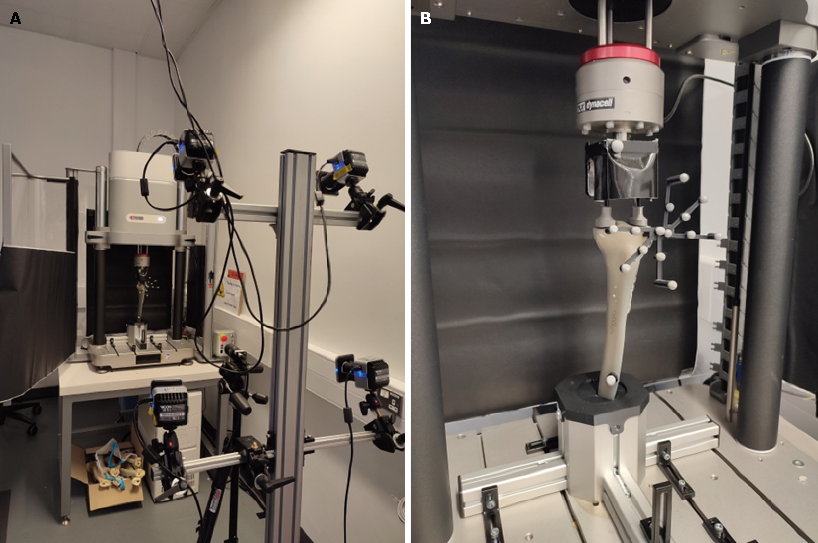
Figure 4 Mechanical testing arrangement.
A: The testing set-up; B: Closeup showing the bespoke support, loading structures and the marker clusters for fracture gap tracking.
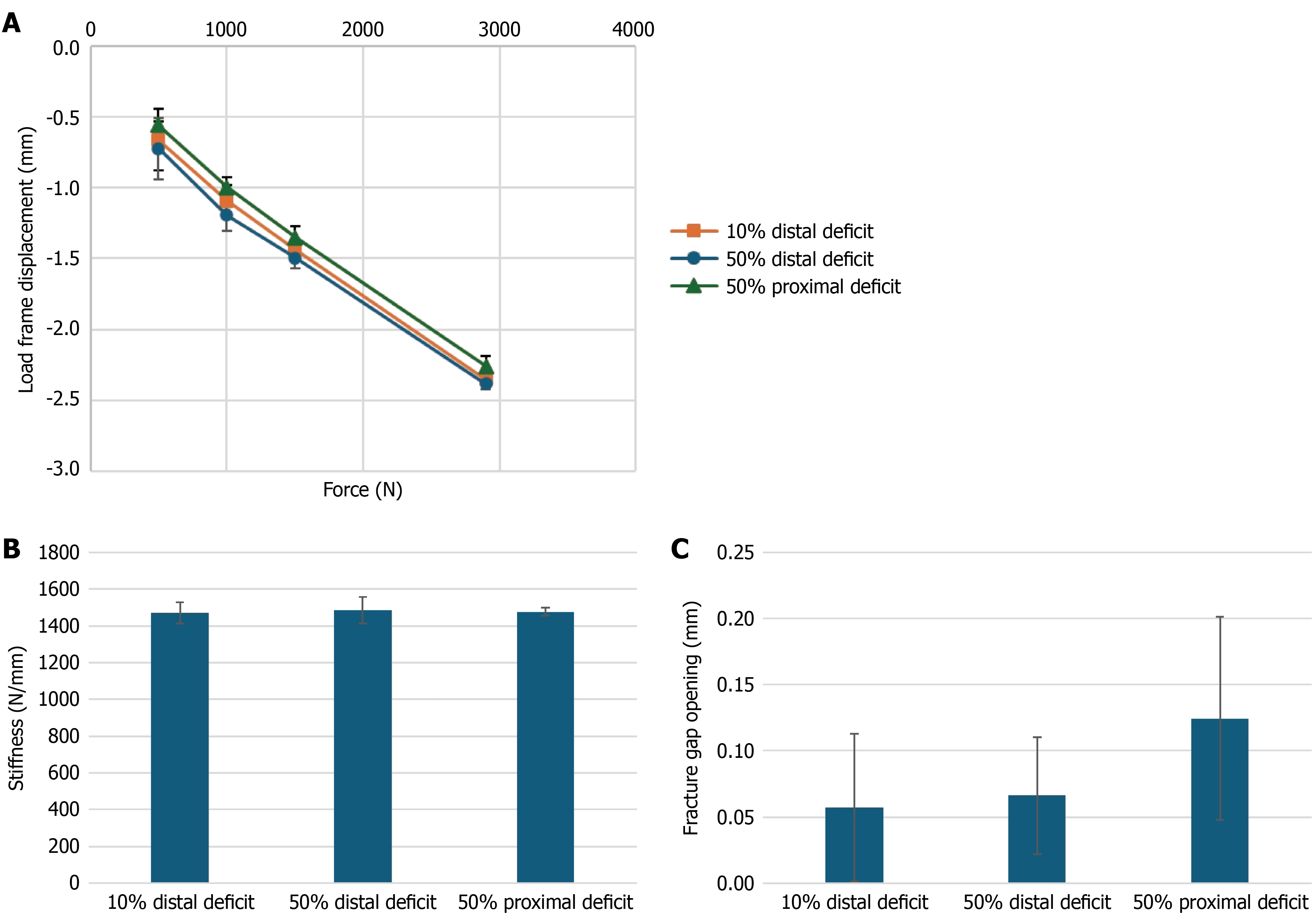
Figure 5 Biomechanical performance across the three cancellous bone defect scenarios.
A: Average vertical displacement for increasing loading; B: Stiffness; C: Fracture gap opening under maximum compression for the three conditions considered.
- Citation: Okoro T, Paavana T, McDonald S, Chatzistergos P, Crooks S, Murugesan T, Kondi S, Needham R, Chockalingam N. Influence of proximal cancellous bone void volume on the rigidity of tibial plateau fracture fixation: An observational study. World J Orthop 2026; 17(11): 123916
- URL: https://www.wjgnet.com/2218-5836/full/v17/i11/123916.htm
- DOI: https://dx.doi.org/10.5312/wjo.123916