Copyright
©The Author(s) 2025.
World J Orthop. Jul 18, 2025; 16(7): 106416
Published online Jul 18, 2025. doi: 10.5312/wjo.v16.i7.106416
Published online Jul 18, 2025. doi: 10.5312/wjo.v16.i7.106416
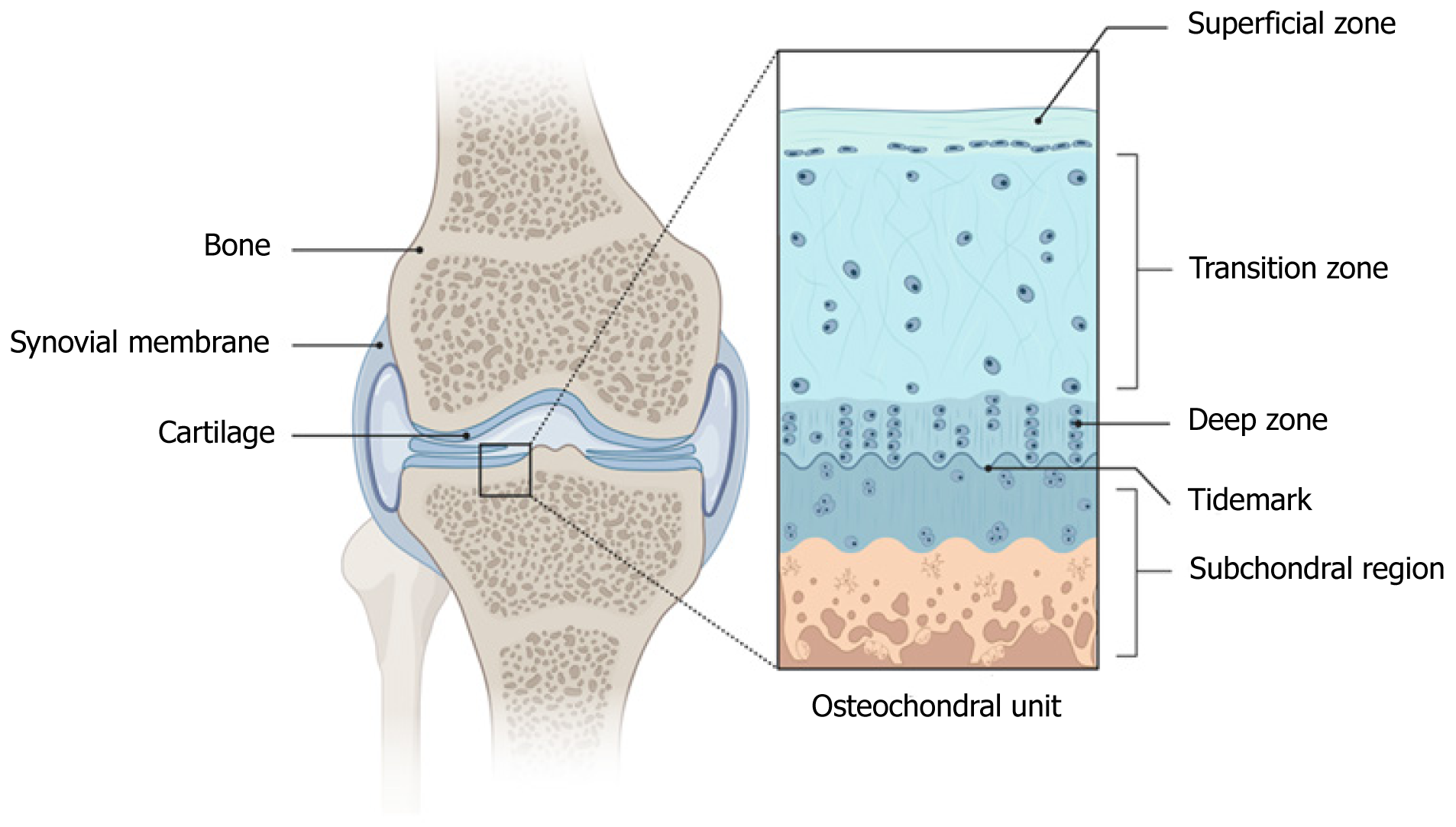
Figure 1 Normal healthy cartilage structure.
Figure 2 Radiograph of the knee cartilage.
A: Conventional; B: Diffraction-enhanced.
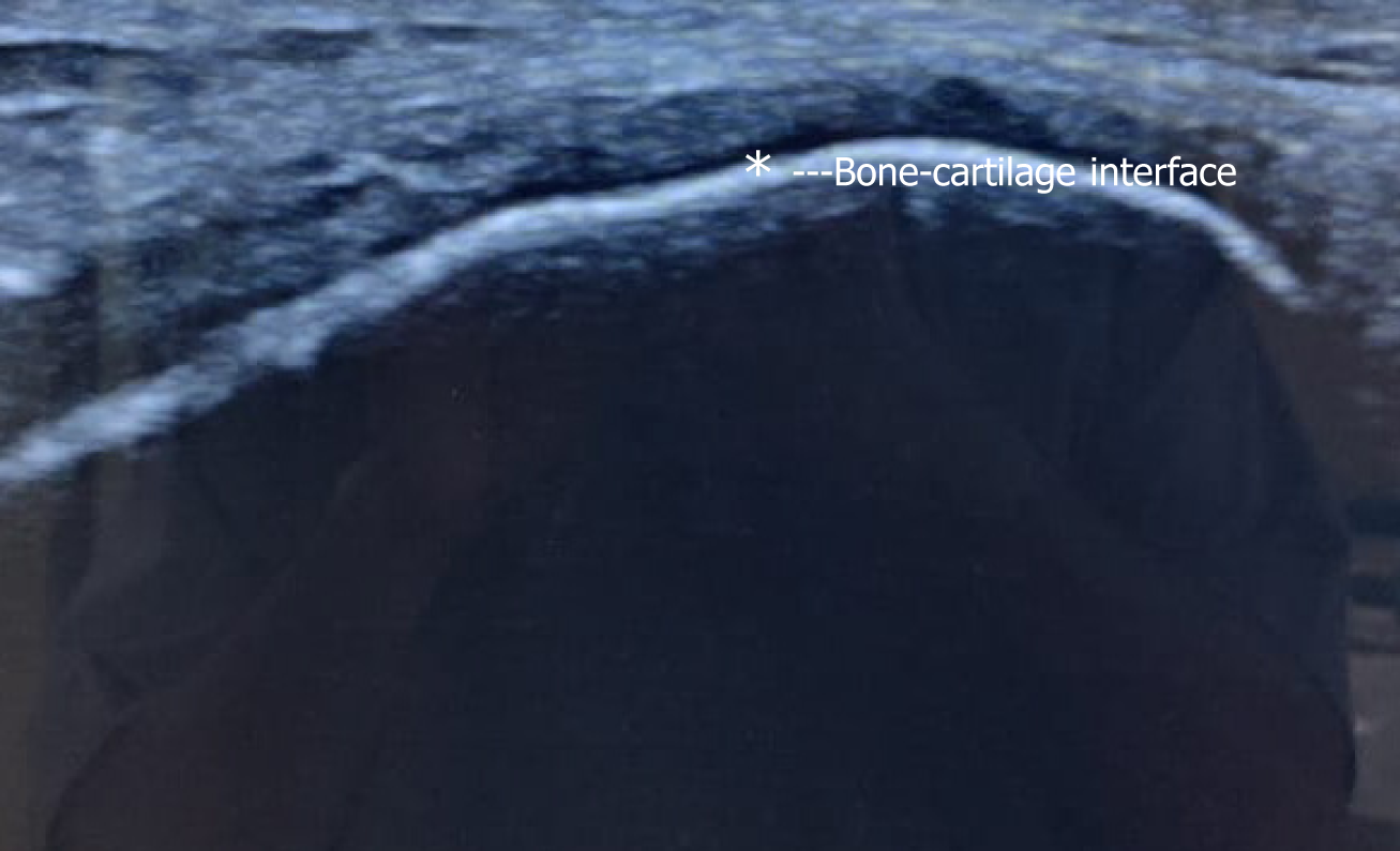
Figure 3 Ultrasound imaging of cartilage.
* indicates hypoechoic cartilage zone.
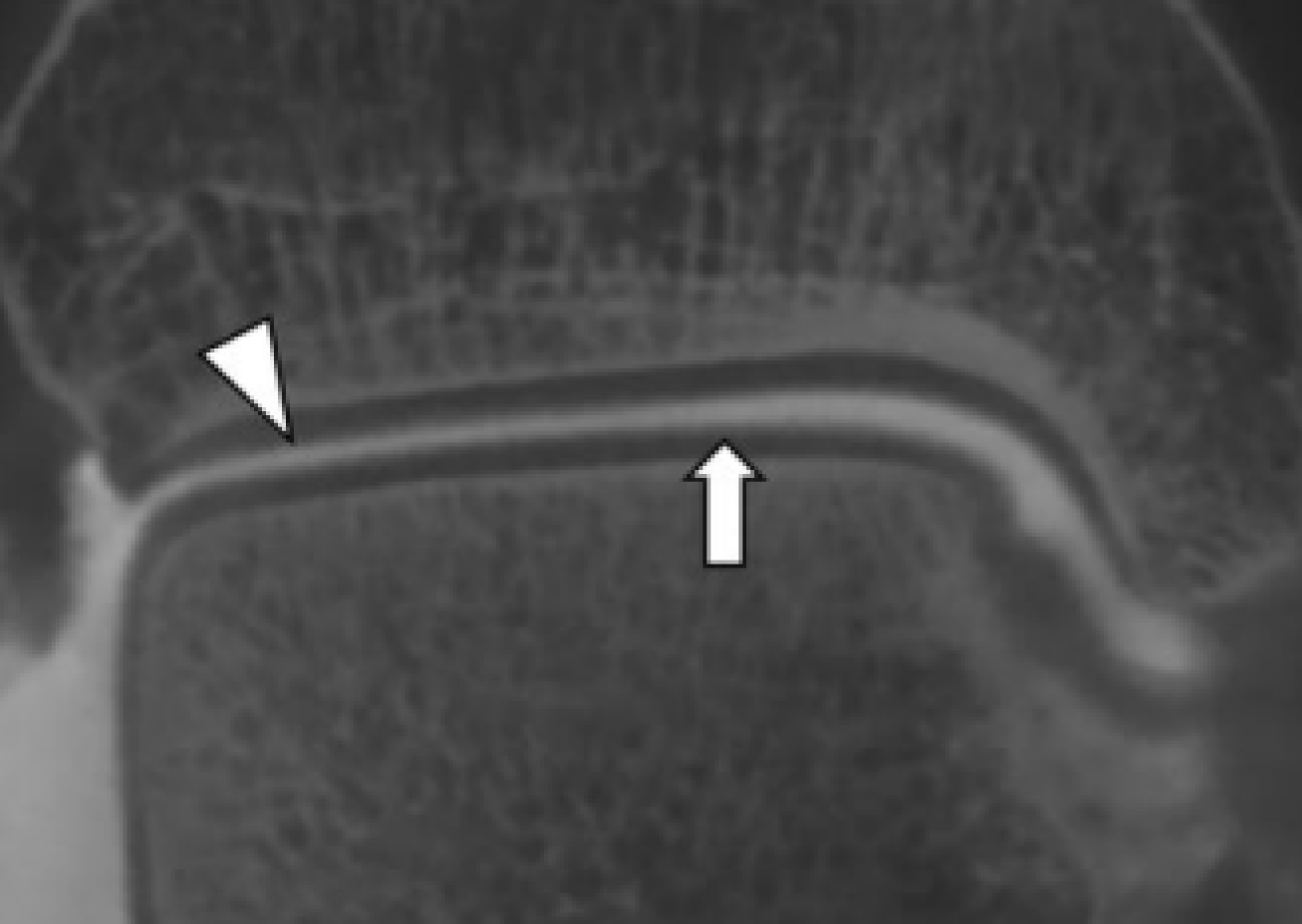
Figure 4 Cone-beam computed tomography arthrogram of tibiotalar joint with an arrowhead showing the contrast in the joint space and arrow marking the distinct margins of the intact cartilage in the talar dome.
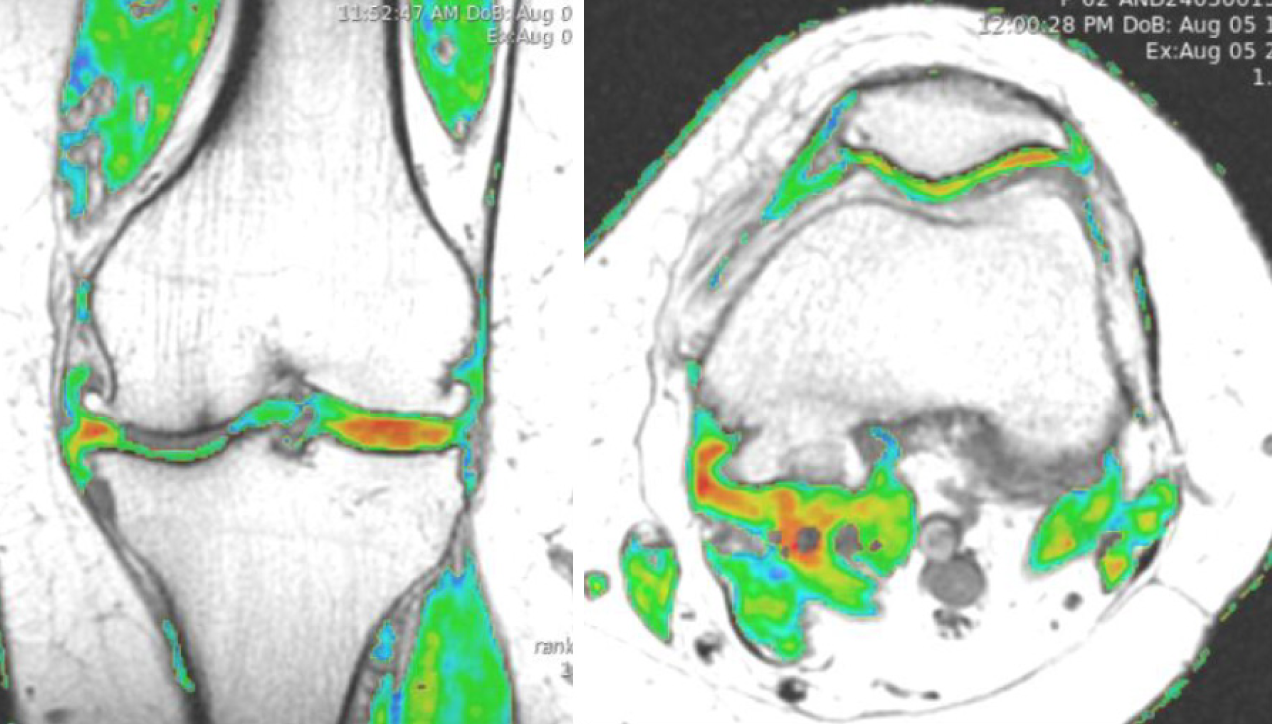
Figure 5 T2-mapping of the magnetic resonance imaging cartigram sequence done to estimate the cartilage degeneration in knee osteoarthritis.
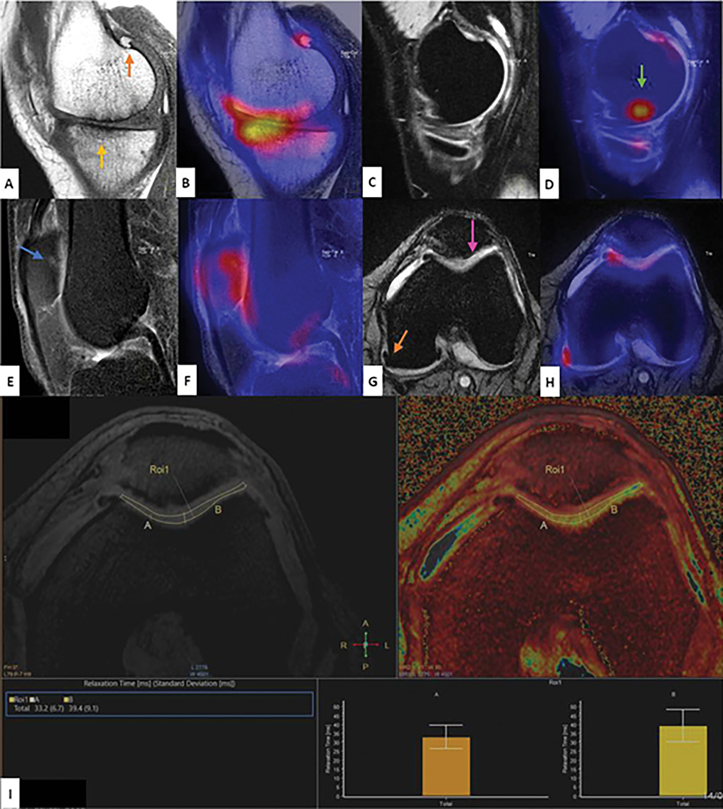
Figure 6 18F sodium fluoride (18F-NaF) served as a one-stop modality to assess whole joint, that is, bone pathology, cartilage, and ligaments (sagittal T1 turbo spin echo) showed osteophyte (orange arrow) and sclerosis (yellow arrow) with corresponding 18F-NaF uptake.
A and B: Fused positron emission tomography/magnetic resonance imaging (PET/MRI); C: Sagittal three-dimensional MEDIC did not have any structural abnormality but when fused with PET; D: There was high uptake volume of interest (white arrow) termed as “subchondral magic spot”; E and F: Sagittal T2 spectral attenuated inversion recovery depicted grade 1 bone marrow lesion (blue arrow) with corresponding uptake in fused PET/MRI; G: Axial T2* showed degenerated medial and lateral trochlear cartilage (pink arrow); H: Corresponding fused PET/MRI; I: T2 relaxometry with raised values. A-I: Citation: Jena A, Goyal N, Rana P, Taneja S, Vaish A, Botchu R, Vaishya R. Qualitative and Quantitative Evaluation of Morpho-Metabolic Changes in Bone Cartilage Complex of Knee Joint in Osteoarthritis Using Simultaneous 18F-NaF PET/MRI-A Pilot Study. Indian J Radiol Imaging 2023; 33: 173-182. Copyright© Indian Radiological Association 2023. Published by Thieme Medical and Scientific Publishers. This is an open access article published by Thieme under the terms of the Creative Commons Attribution-NonDerivative-NonCommercial License, permitting copying and reproduction so long as the original work is given appropriate credit. Contents may not be used for commercial purposes, or adapted, remixed, transformed or built upon (https://creativecommons.org/licenses/by-nc-nd/4.0/).
- Citation: Jeyaraman M, Jeyaraman N, Nallakumarasamy A, Ramasubramanian S, Muthu S. Insights of cartilage imaging in cartilage regeneration. World J Orthop 2025; 16(7): 106416
- URL: https://www.wjgnet.com/2218-5836/full/v16/i7/106416.htm
- DOI: https://dx.doi.org/10.5312/wjo.v16.i7.106416


