Copyright
©The Author(s) 2023.
World J Orthop. May 18, 2023; 14(5): 328-339
Published online May 18, 2023. doi: 10.5312/wjo.v14.i5.328
Published online May 18, 2023. doi: 10.5312/wjo.v14.i5.328
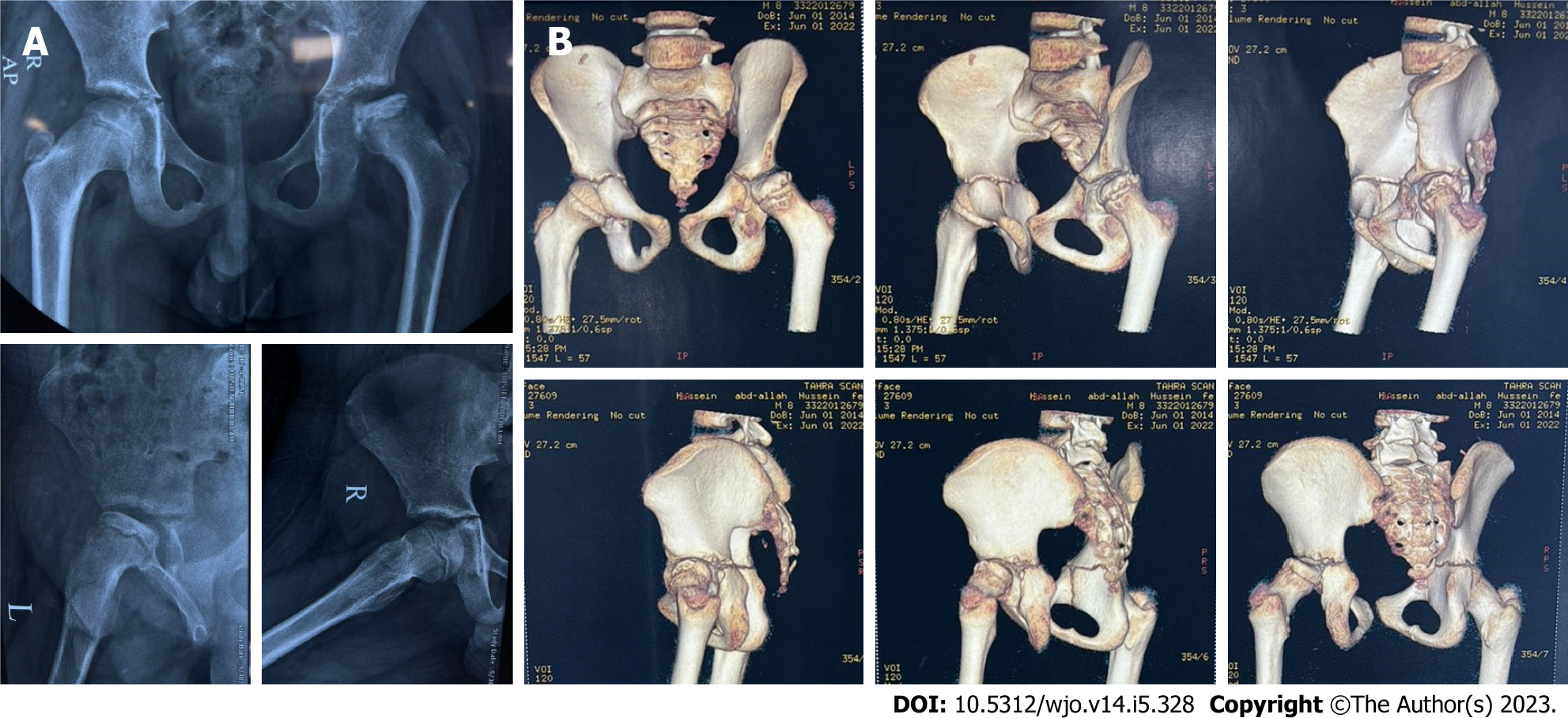
Figure 1 The extent of limb deformity.
A: Preoperative Plain X ray of 11 years male patient with left Perthes disease (Waldenström: Healing stage); B: Preoperative 3D-computed tomography scan.
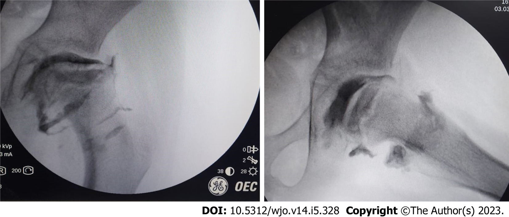
Figure 2 Intraoperative arthrography of the previous patient showing that the head is aspherical with lateral osteophytes.
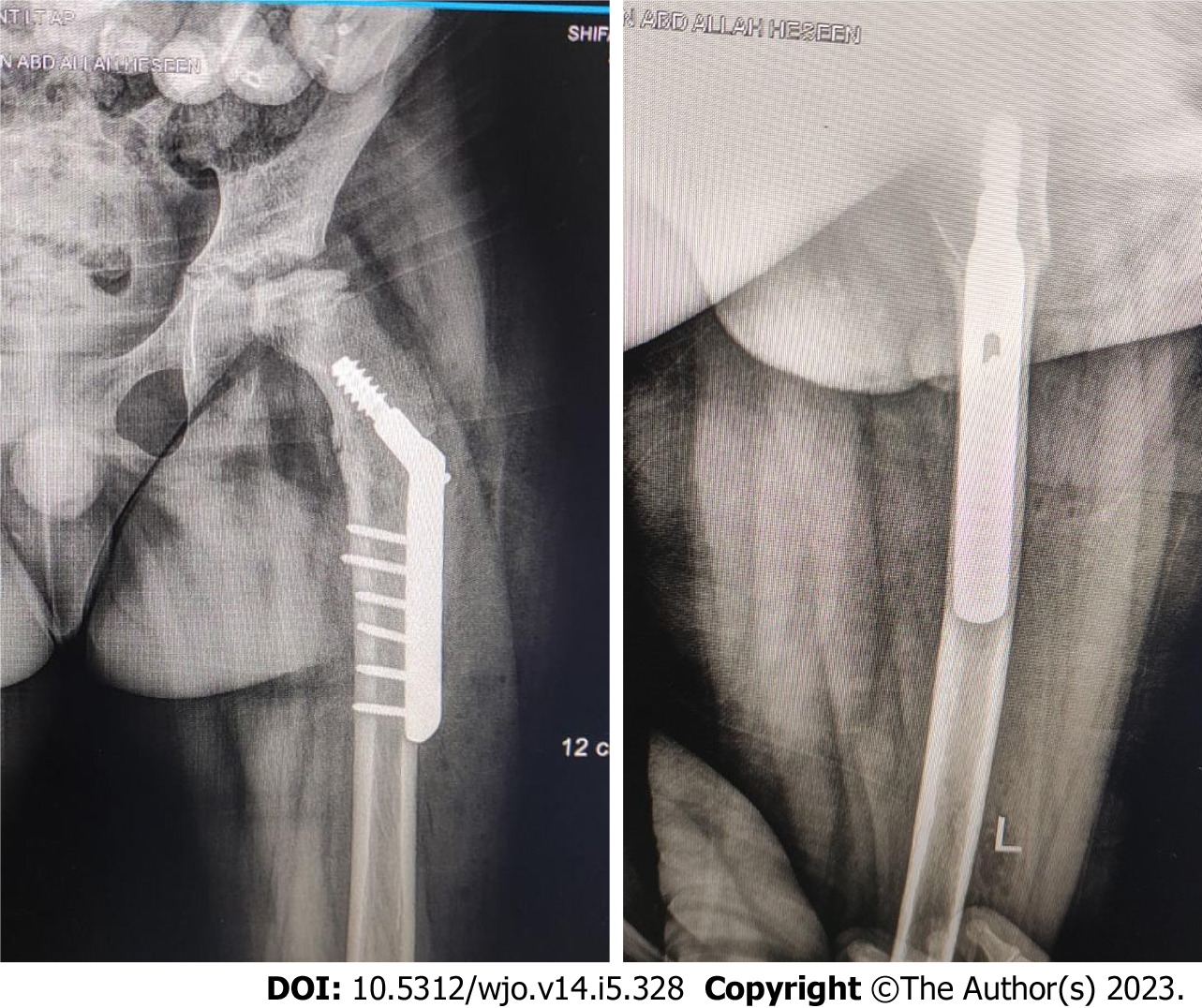
Figure 3 Immediate post operative pregnane X receptor of the previous patient fixed with dynamic hip screw.
10° valgus, 10° extension and 15° internal rotation were done.
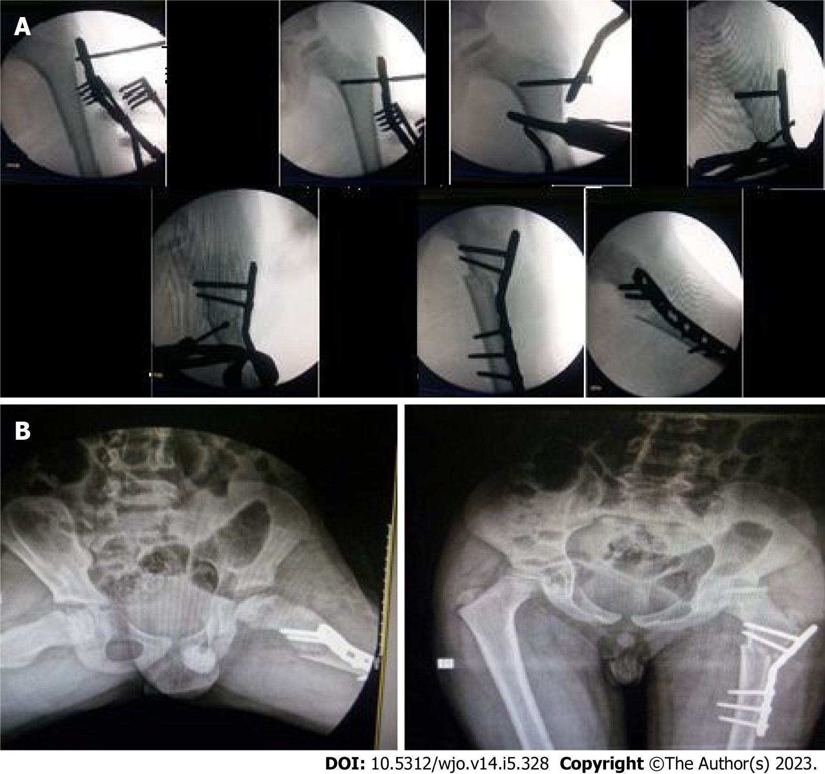
Figure 4 Treatment of a femoral valgus osteotomy in a 10-year-old man with left Pertes disease.
A: Intra-operative surgical steps of another 10 years old male patient with left Perthes disease (Waldenström: Re-ossification stage) treated with femoral valgus osteotomy using pre-contoured plate. Note the extension component of the osteotomy was evident in the lateral shot to compensate associated fixed flexion deformity of the hip; B: Immediate post operative pelvic X-ray of the same patient.
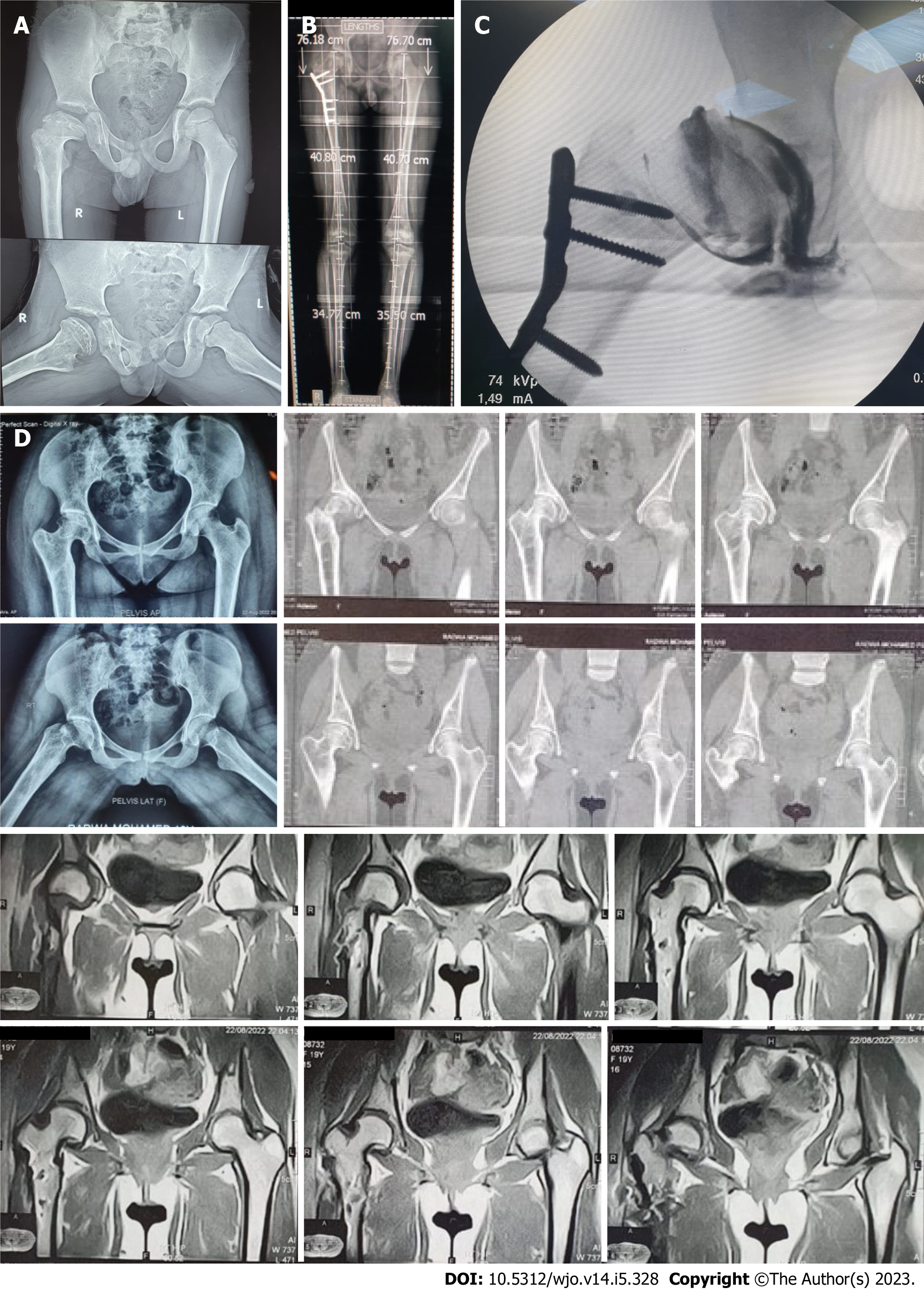
Figure 5 Clinical evaluation for 8-11 years in patients with femoral valgus osteotomy with a rotational component.
A: Preoperative pelvic X-ray (PXR) of 9 years old male patient with right Perthes disease (Waldenström: Healing stage); B: Follow up scanogram after 10 years showing no limb length discrepancy nor hip arthritis; C: Intraoperative hip arthrography just before plate removal that show smooth head with no osteophytes; D: Post removal PXR, computed tomography and magnetic resonance imaging respectively that show normal hip.
- Citation: Emara KM, Diab RA, Emara AK, Eissa M, Gemeah M, Mahmoud SA. Mid-term results of sub-trochanteric valgus osteotomy for symptomatic late stages Legg-Calvé-Perthes disease. World J Orthop 2023; 14(5): 328-339
- URL: https://www.wjgnet.com/2218-5836/full/v14/i5/328.htm
- DOI: https://dx.doi.org/10.5312/wjo.v14.i5.328


