Copyright
©The Author(s) 2023.
World J Orthop. May 18, 2023; 14(5): 302-311
Published online May 18, 2023. doi: 10.5312/wjo.v14.i5.302
Published online May 18, 2023. doi: 10.5312/wjo.v14.i5.302
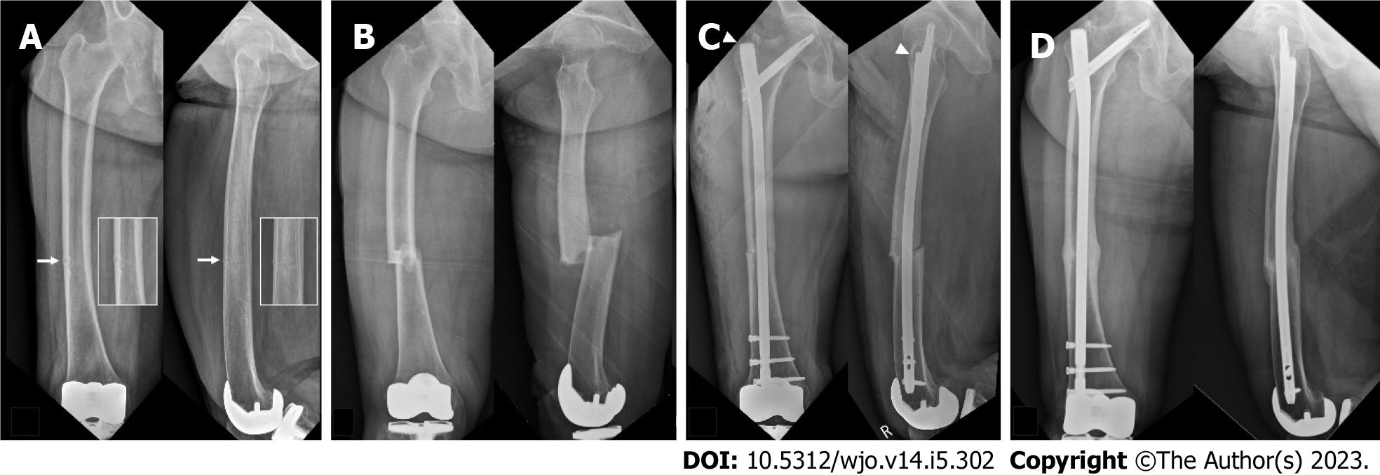
Figure 1 Images of an 80-year-old woman.
A: An 80-year-old woman was transferred from a spine clinic because of intractable right thigh pain for three months. Radiographs revealed a transverse radiolucent line (white arrows and insets) on the lateral and anterior cortex of the right femur with 10° of varus. The patient refused prophylactic surgery for incomplete atypical femoral fracture (AFF); however, medical treatment including a switch from bisphosphonate to teriparatide was initiated. According to a scoring system[11], the risk for impending complete AFF was scored as 10 points; B: Two months later, she visited the emergency department due to progression to a complete AFF; C: She underwent fixation with a long cephalomedullary nail (Trochanteric Fixation Nail-Advanced®, DePuy Synthes, Winterthur, Switzerland) spanning the whole length of the femur. It is to be noted that the entry point of the nail is lateral and anterior to the greater trochanter tip (arrowheads); D: Radiographs taken at 18 mo postoperatively showed healing of the fracture site.
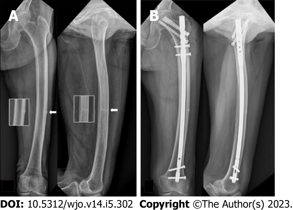
Figure 2 Images of an 81-year-old woman who had taken bisphosphonate for two years.
A: An 81-year-old woman who had taken bisphosphonate for two years visited the outpatient clinic complaining of left thigh pain for two months. Radiographs revealed a transverse beak and radiolucent line (white arrows) on the lateral and anterior cortex of the right femur with 10° of varus and 7° of anterior angulation. According to a scoring system[11], the risk for impending complete atypical femoral fracture was scored as 11 points; B: She underwent fixation with an opposite-side (right side) standard nail (Sirus Femoral Nail®, Zimmer, Warsaw, IN, USA). Prophylactic screw fixation toward the femoral neck on her left femur was performed to prevent potential femoral neck fracture around the nail.
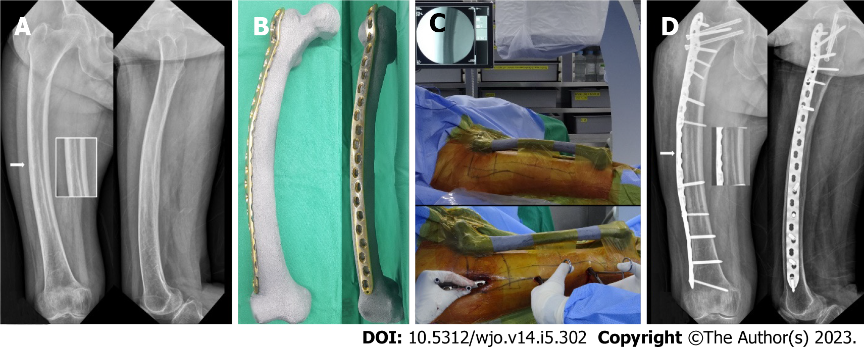
Figure 3 Images of a 75-year-old woman underwent a fixation with a long standard nail.
A: A 75-year-old woman underwent a fixation with a long standard nail due to a complete atypical femoral fracture (AFF) on her right femur two years ago at another hospital and kept taking bisphosphonate (BP) until she visited our clinic; B: She reported left groin and thigh pain for six months. Radiographs revealed arthritis on her left hip joint (arrowheads) and transverse beaks with radiolucent lines (“dreaded black line”) on the lateral and anterior cortex of the left femur (white arrows and insets). According to a scoring system[11], the risk for impending complete AFF was scored as 8 points and BP medication was discontinued; C: Before total hip arthroplasty (THA) to treat hip arthritis, a locking compression plate was pre-contoured along the shape of the bone model with 3D printing rapid prototyping; D: During THA, the sterile 3D-printed model was placed in the same position as that of the femur and used as a surgical navigation. Fixation with the pre-contoured plate via minimally invasive plate osteosynthesis was performed to treat incomplete AFF; E: Radiographs taken three years postoperatively showed complete healing of the AFF without progression of femoral bowing or implant-related complications.
Figure 4 Images of a 75-year-old woman who had taken bisphosphonate.
A: A 75-year-old woman who had taken bisphosphonate for a period of four years visited our clinic with right thigh pain for three months. Radiographs showed a transverse radiolucent line (white arrow and inset) on the apex of the lateral cortex of the right femur with 7° of varus. According to a scoring system[11], the risk for impending complete atypical femoral fracture (AFF) was scored as 9 points; B: Before the surgery, a locking compression plate was pre-contoured along the shape of the bone model with 3D printing rapid prototyping; C: During the surgery, the sterile 3D-printed bone model was placed in the same position as that of the femur and used as a surgical navigation; D: Fixation with pre-contoured plate fixation for incomplete AFF (white arrow and inset) with severe bowing was performed via minimally invasive plate osteosynthesis. It is to be noted that additional prophylactic screw fixation toward the femoral neck was performed to prevent potential femoral neck fractures.
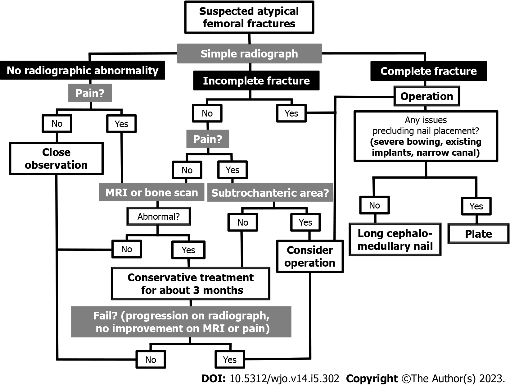
Figure 5 A proposed treatment algorithm for suspected atypical femoral fracture.
MRI: Magnetic resonance imaging.
- Citation: Shim BJ, Won H, Kim SY, Baek SH. Surgical strategy of the treatment of atypical femoral fractures. World J Orthop 2023; 14(5): 302-311
- URL: https://www.wjgnet.com/2218-5836/full/v14/i5/302.htm
- DOI: https://dx.doi.org/10.5312/wjo.v14.i5.302


