Copyright
©The Author(s) 2023.
World J Orthop. Apr 18, 2023; 14(4): 260-267
Published online Apr 18, 2023. doi: 10.5312/wjo.v14.i4.260
Published online Apr 18, 2023. doi: 10.5312/wjo.v14.i4.260
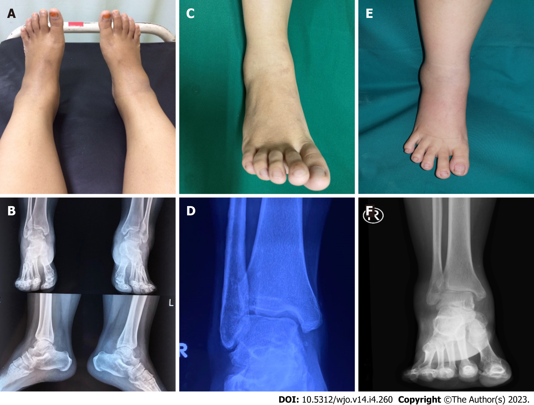
Figure 1 Clinical and radiological images of case 1, 2, and 3, respectively.
A, B: Case 1; C, D: Case 2; E, F: Case 3.
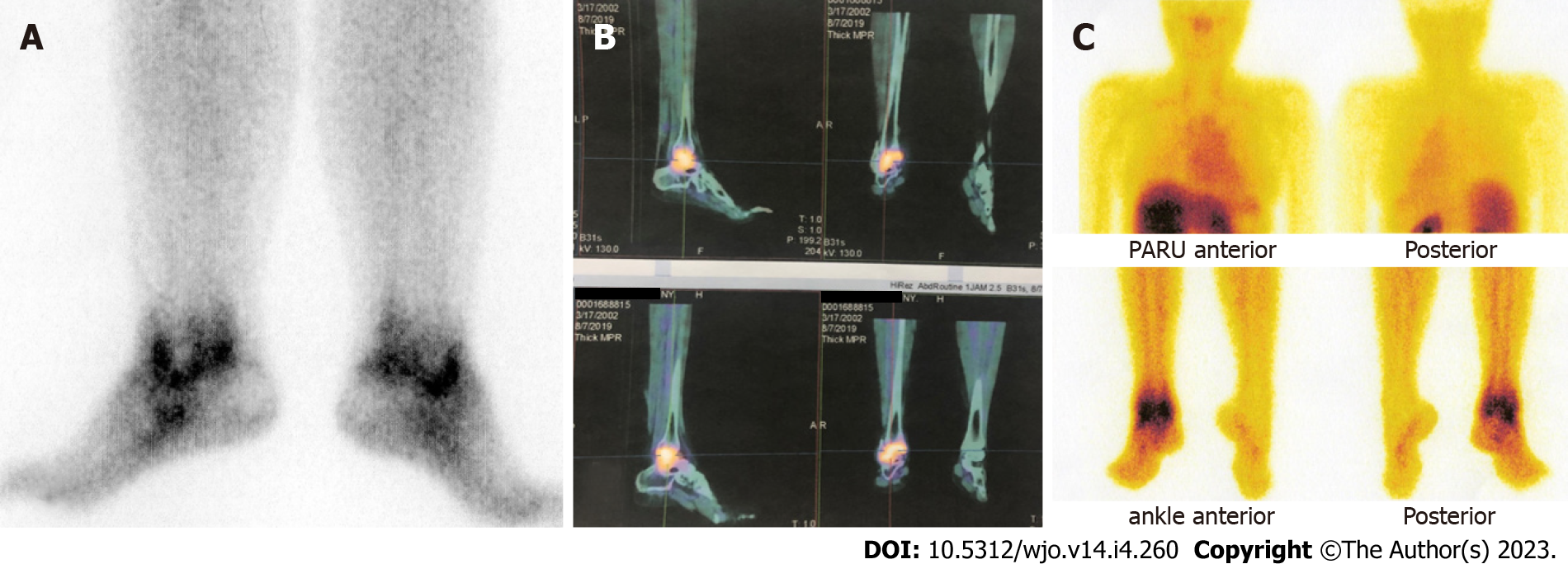
Figure 2 Scintigraphy images of case 1, 2, and 3, respectively.
A: Case 1; B: Case 2; C: Case 3.
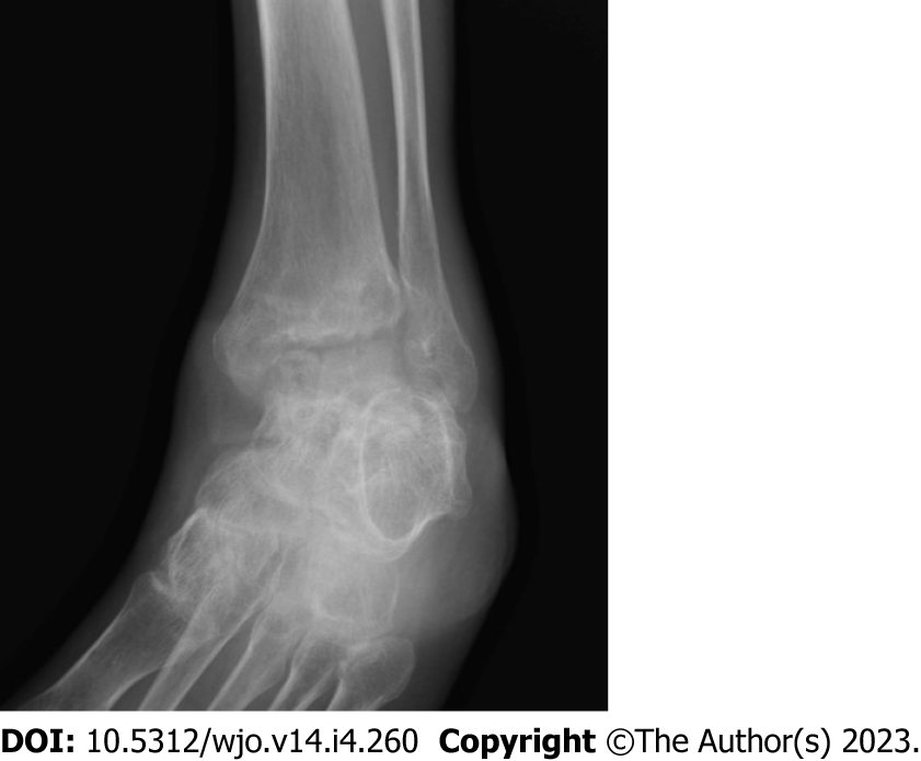
Figure 3 Radiological image depicting Phemister Triad.
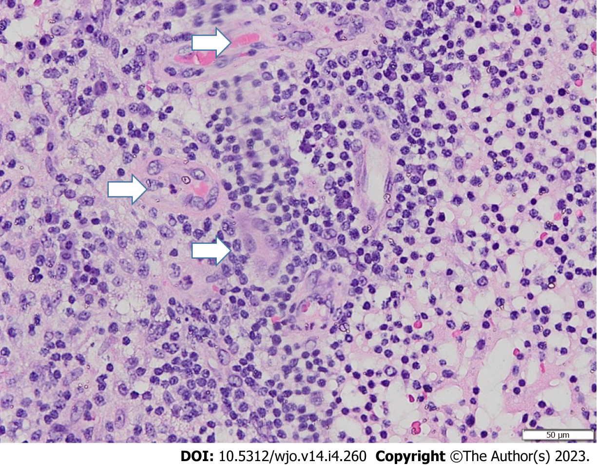
Figure 4 Histopathological image showing multinucleated giant cells (arrows).
- Citation: Primadhi RA, Kartamihardja AHS. Subclinical ankle joint tuberculous arthritis - The role of scintigraphy: A case series. World J Orthop 2023; 14(4): 260-267
- URL: https://www.wjgnet.com/2218-5836/full/v14/i4/260.htm
- DOI: https://dx.doi.org/10.5312/wjo.v14.i4.260


