Copyright
©The Author(s) 2023.
World J Orthop. Apr 18, 2023; 14(4): 171-185
Published online Apr 18, 2023. doi: 10.5312/wjo.v14.i4.171
Published online Apr 18, 2023. doi: 10.5312/wjo.v14.i4.171
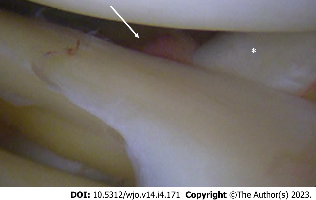
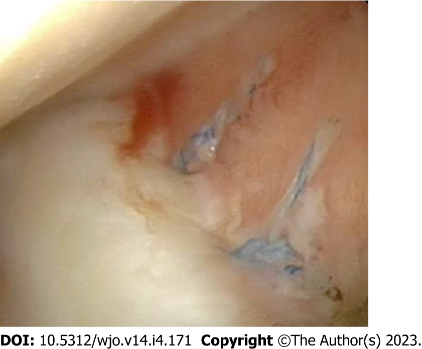
Figure 1 Arthroscopic view of posterosuperior popliteomeniscal fascicule injury (white arrow).
The lateral meniscus is no longer properly anchored to popliteus tendon (asterisk), causing meniscus hypermobility.
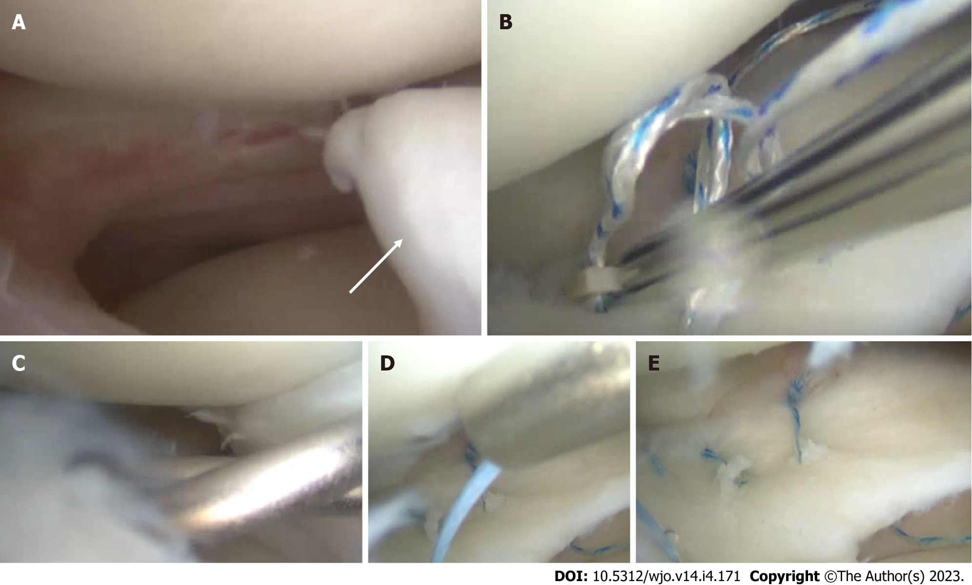
Figure 2 Isolated lateral meniscus bucket-handle tear in an 18-year-old male patient.
A: Arthroscopic findings with the unstable bucket-handle fragment (white arrow); B: Arthroscopic repair of the posterior horn using an all-inside technique; C: Repair of the anterior third using an outside-in technique; D: Repair of the meniscal body with an inside-out technique; E: Final aspect of the meniscal repair.
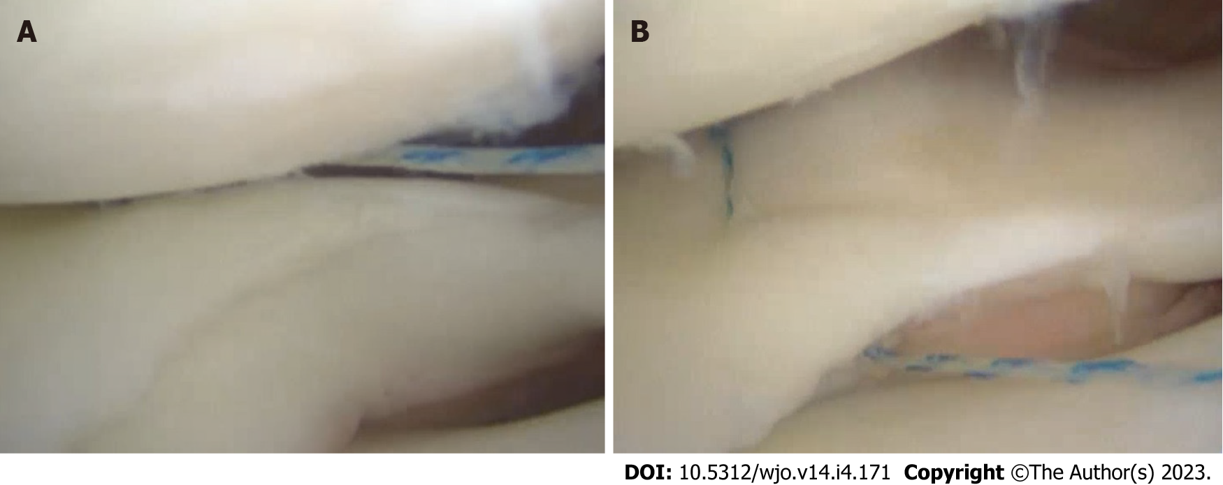
Figure 3 All-inside repair of a longitudinal meniscus tear.
A: Superior suture placement creates a non-uniform compression of the tear; B: A second suture placed in the inferior portion of the meniscus rebalance the repair compression forces, thus correcting the superior meniscus displacement secondary to the first suture.
Figure 4 Ramp lesion repair from the posteromedial portal using a suture hook.
A: The suture is passed through the posteromedial capsule and the posterior meniscotibial ligament; B: The hook is advanced until reaching the posterior portion of the medial meniscus; C: A sliding-knot is used to secure the repair (view from the posteromedial portal).
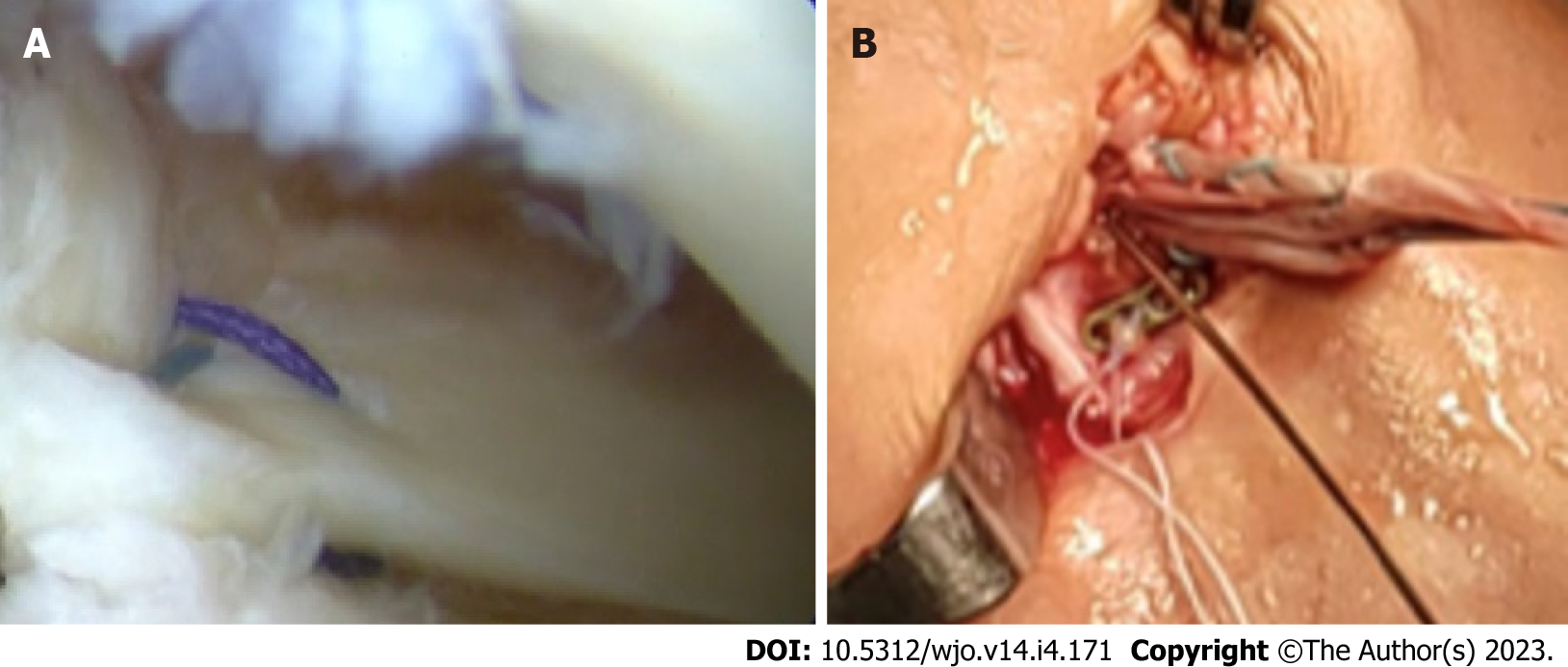
Figure 5 Arthroscopic repair of the lateral meniscus posterior root repair.
A: Arthroscopic view of the repair; B: The suture is passed in the same anterior cruciate ligament tibial tunnel and knotted on a dedicated plate.
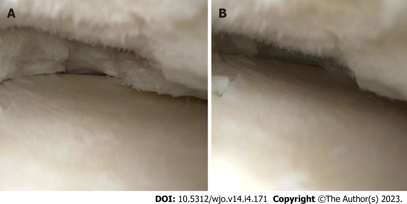
Figure 6 Arthroscopic view of a radial tear of the medial meniscus.
A: Medial meniscus in no-weightbearing condition; B: After applying a compression force, the medial meniscus is totally extruded, thus losing is protective function in the knee joint.
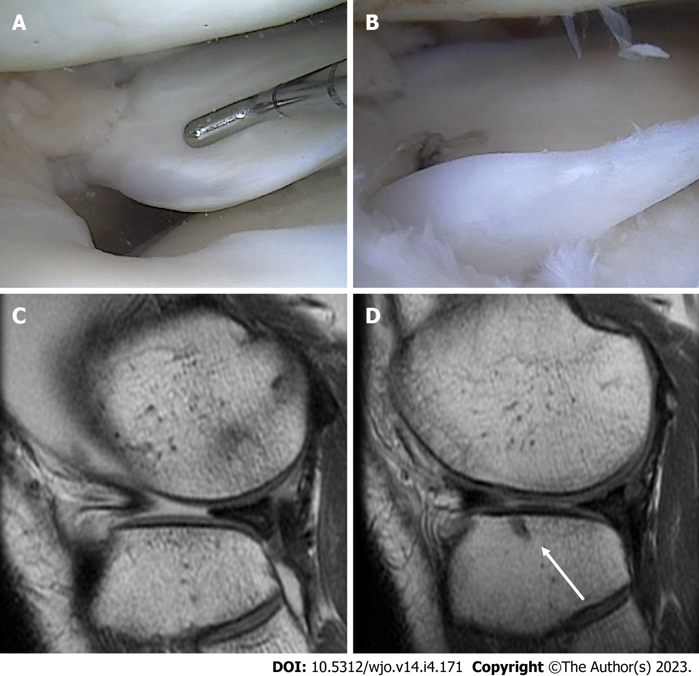
Figure 7 Radial tear of the lateral meniscus.
A: Arthroscopic visualization of the radial tear of the lateral meniscus; B: Tear repair using an all-suture anchor; C: Preoperative T1 sagittal magnetic resonance imaging (MRI) view, showing the extrusion of the anterior portion of the lateral meniscus; D: Six-month follow-up T1 sagittal MRI view, showing the relocation of the anterior portion and the all-suture anchor used for the repair (white arrow).
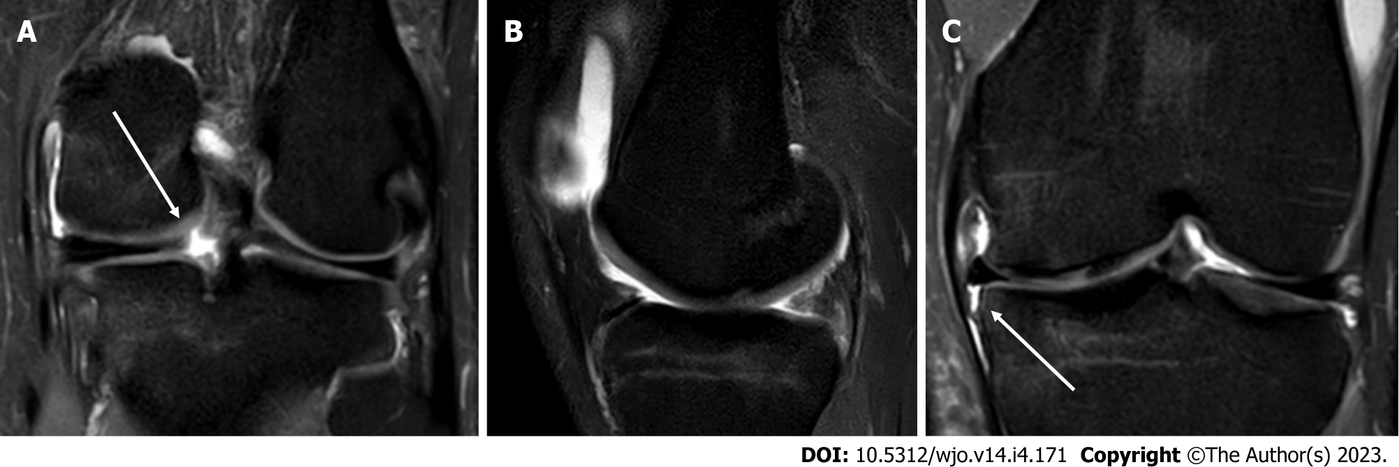
Figure 8 Isolated posterior root tear of the medial meniscus.
A: T2 Fat-Sat coronal magnetic resonance imaging (MRI) view, showing disruption of the meniscus ring (white arrow); B: T2 Fat-Sat sagittal MRI view, with the “ghost sign”, highly suggestive for posterior root tear; C: T2 Fat-Sat coronal MRI view, showing detachment of the medial meniscotibial ligament (white arrow) and meniscus extrusion.
Figure 9 Arthroscopic view of medial meniscus centralization with two all-suture anchors.
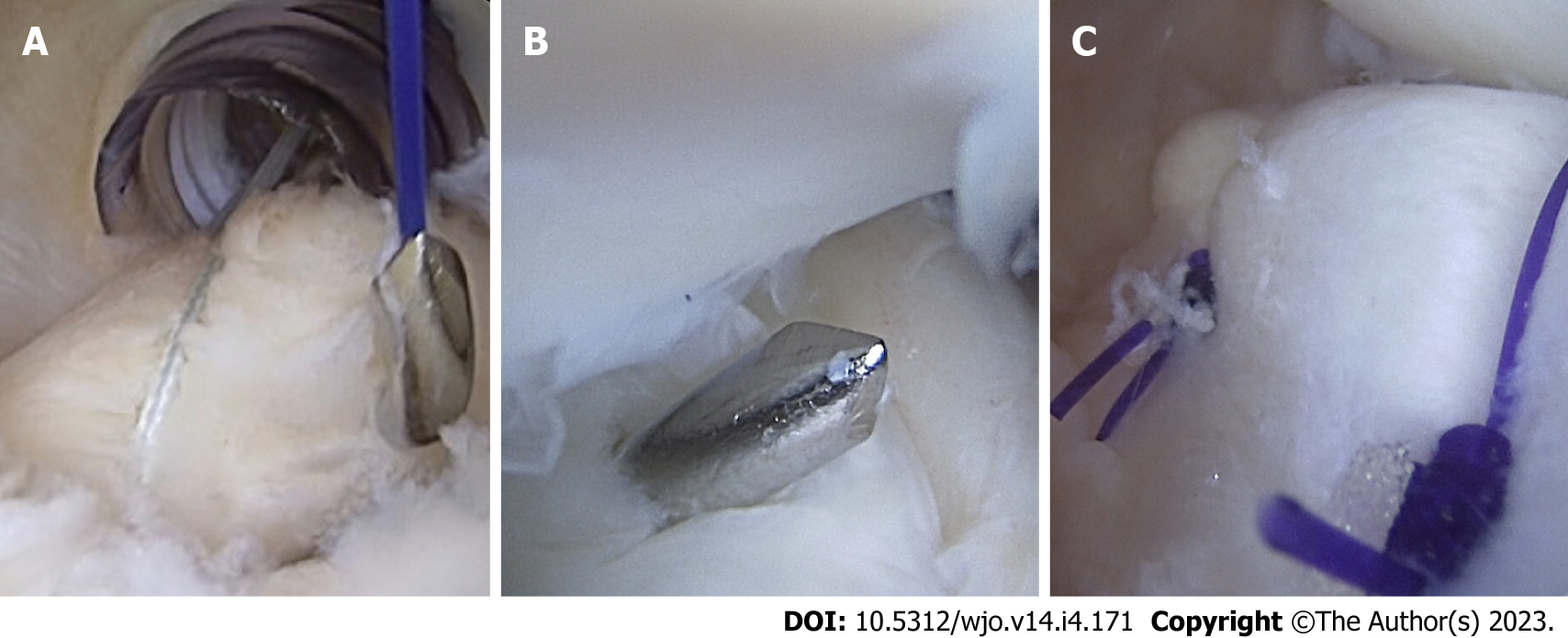
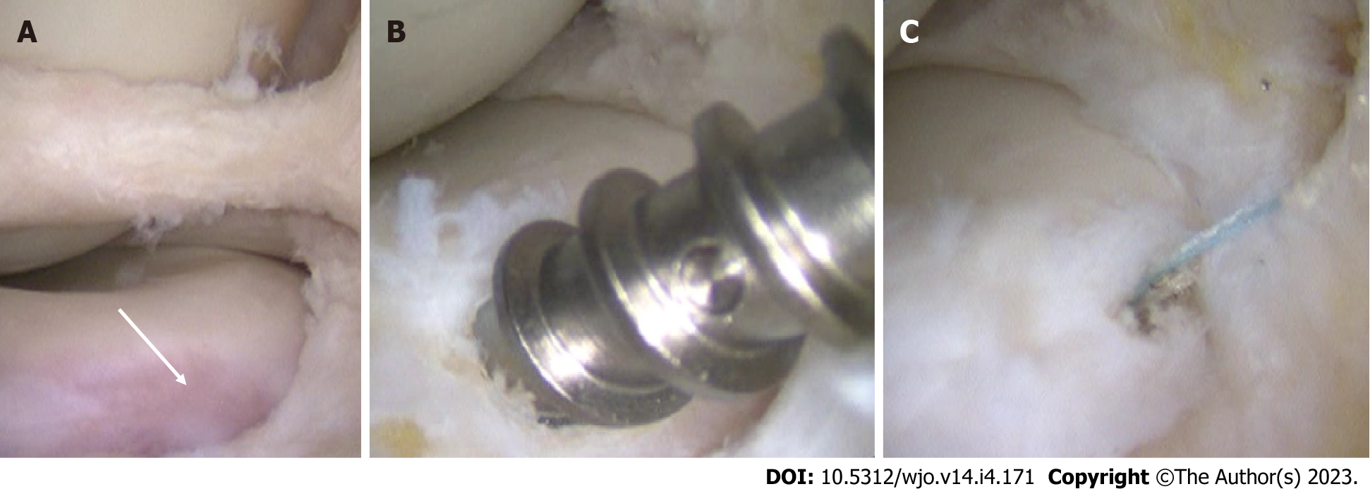
Figure 10 Anterior root detachment of the medial meniscus, with concomitant bucket handle tear.
A: Arthroscopic finding with the tibial avulsion site (white arrow); B: Repair of the lesion using a 3.5 metallic suture anchor; C: Final view of the repair.
- Citation: Simonetta R, Russo A, Palco M, Costa GG, Mariani PP. Meniscus tears treatment: The good, the bad and the ugly-patterns classification and practical guide. World J Orthop 2023; 14(4): 171-185
- URL: https://www.wjgnet.com/2218-5836/full/v14/i4/171.htm
- DOI: https://dx.doi.org/10.5312/wjo.v14.i4.171


