Copyright
©The Author(s) 2022.
World J Orthop. Jan 18, 2022; 13(1): 36-57
Published online Jan 18, 2022. doi: 10.5312/wjo.v13.i1.36
Published online Jan 18, 2022. doi: 10.5312/wjo.v13.i1.36
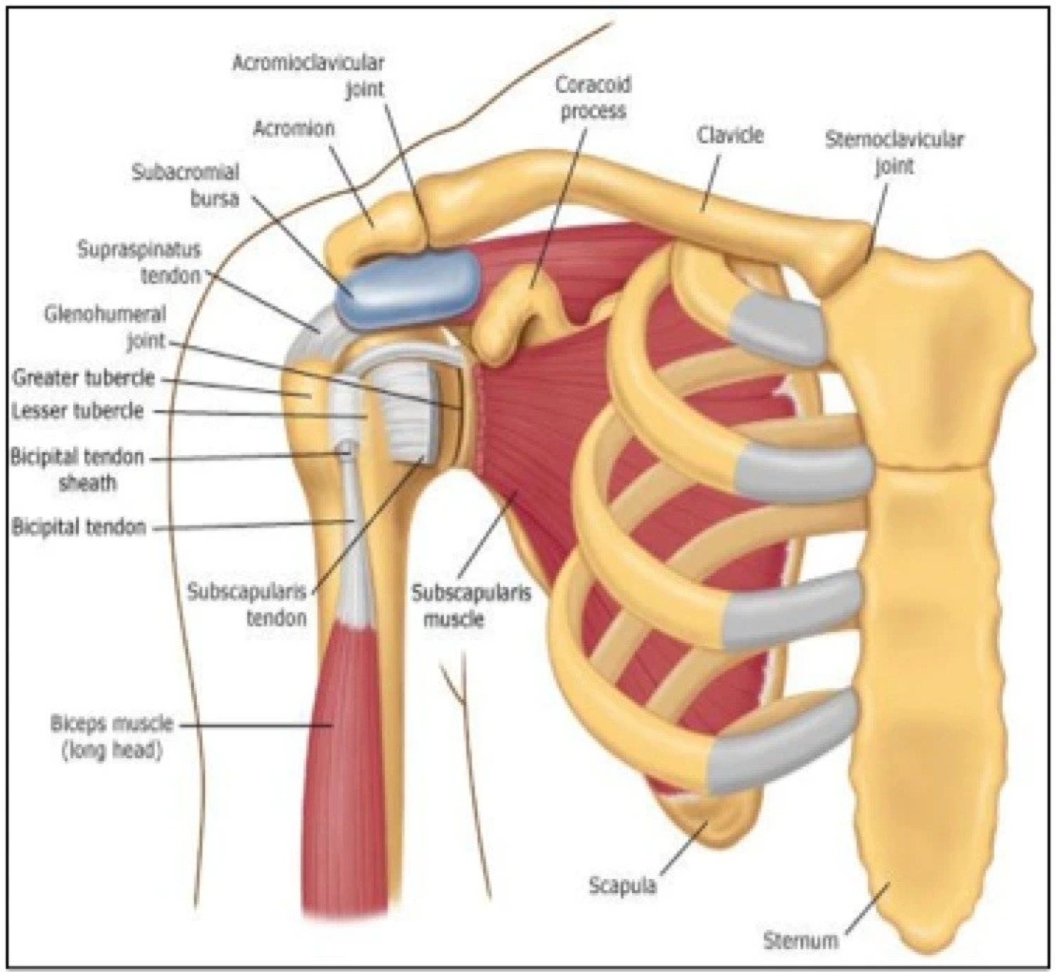
Figure 1 Schematic illustration of anterior shoulder anatomy from Blum et al[9].
Citation: Blum K, Chen AL, Chen TJ, Waite RL, Downs BW, Braverman ER, Kerner MM, Savarimuthu SM, DiNubile N. Repetitive H-wave device stimulation and program induces significant increases in the range of motion of post operative rotator cuff reconstruction in a double-blinded randomized placebo controlled human study. BMC Musculoskelet Disord 2009; 10: 132. Copyright© The Authors 2009. Published by BioMed Central Ltd. This is an open access article distributed under the terms of the Creative Commons CC BY license, which permits unrestricted use, distribution, and reproduction in any medium, provided the original work is properly cited.
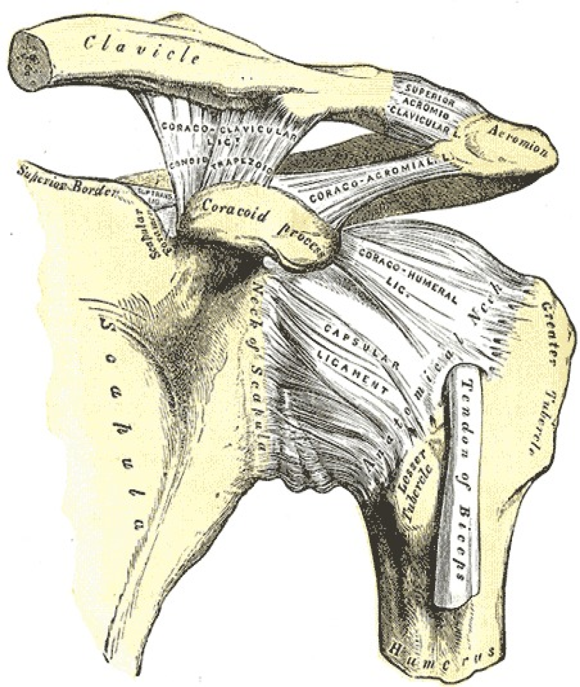
Figure 2 Anterior view of the left shoulder joint depicting tendons and ligaments from Miniato et al[10].
Citation: Miniato MA, Anand P, Varacallo M. Anatomy, Shoulder and Upper Limb, Shoulder. [Updated 2020 Jul 31]. In: StatPearls [Internet]. Treasure Island (FL): StatPearls Publishing. Available from: https://www.ncbi.nlm.nih.gov/books/NBK536933/. Copyright© The Authors 2021. Published by StatPearls Publishing LLC. This book is distributed under the terms of the Creative Commons Attribution 4.0 International License, which permits use, duplication, adaptation, distribution, and reproduction in any medium or format, as long as you give appropriate credit to the original author(s) and the source, a link is provided to the Creative Commons license, and any changes made are indicated.
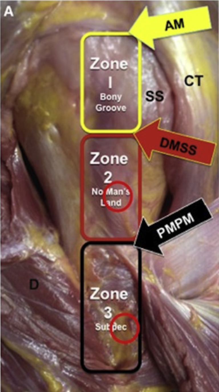
Figure 3 Visual depiction of biceps-labral complex with zone 2 red circle as site for arthroscopic suprapectoral tenodesis and zone 3 red circle as open subpectoral tenodesis location from Forsythe et al[120].
Citation: Forsythe B, Zuke WA, Agarwalla A, Puzzitiello RN, Garcia GH, Cvetanovich GL, Yanke AB, Verma NN, Romeo AA. Arthroscopic Suprapectoral and Open Subpectoral Biceps Tenodeses Produce Similar Outcomes: A Randomized Prospective Analysis. Arthroscopy 2020; 36: 23-32. Copyright© The Authors 2020. Published by Elsevier. The authors have obtained the permission for figure (Supplementary material). AM: Articular margin; CT: Conjoined tendon; d: Deltoid; DMSS: Distal margin of subscapularis tendon; PMPM: Proximal margin of pectoralis major; SS: Subscapularis.
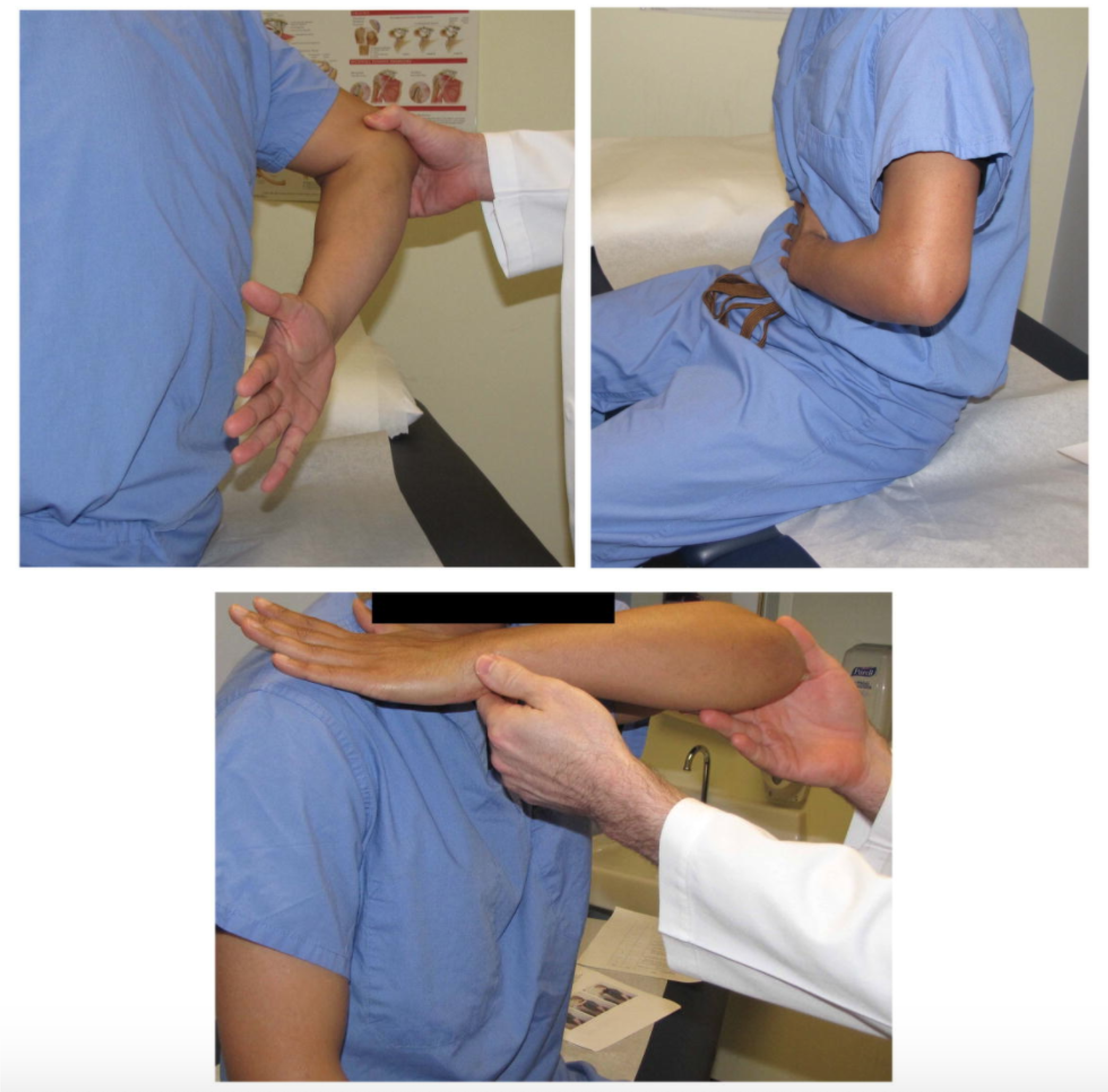
Figure 4 Special tests for subscapularis from Jain et al[47].
Citation: Jain NB, Wilcox RB 3rd, Katz JN, Higgins LD. Clinical examination of the rotator cuff. PM R 2013; 5: 45-56. Copyright© The Authors 2013. Published by John Wiley and Sons. The authors have obtained the permission for figure (Supplementary material). Top left: Lift-off test; Top right: Belly-press test; Bottom: Bear hug test.
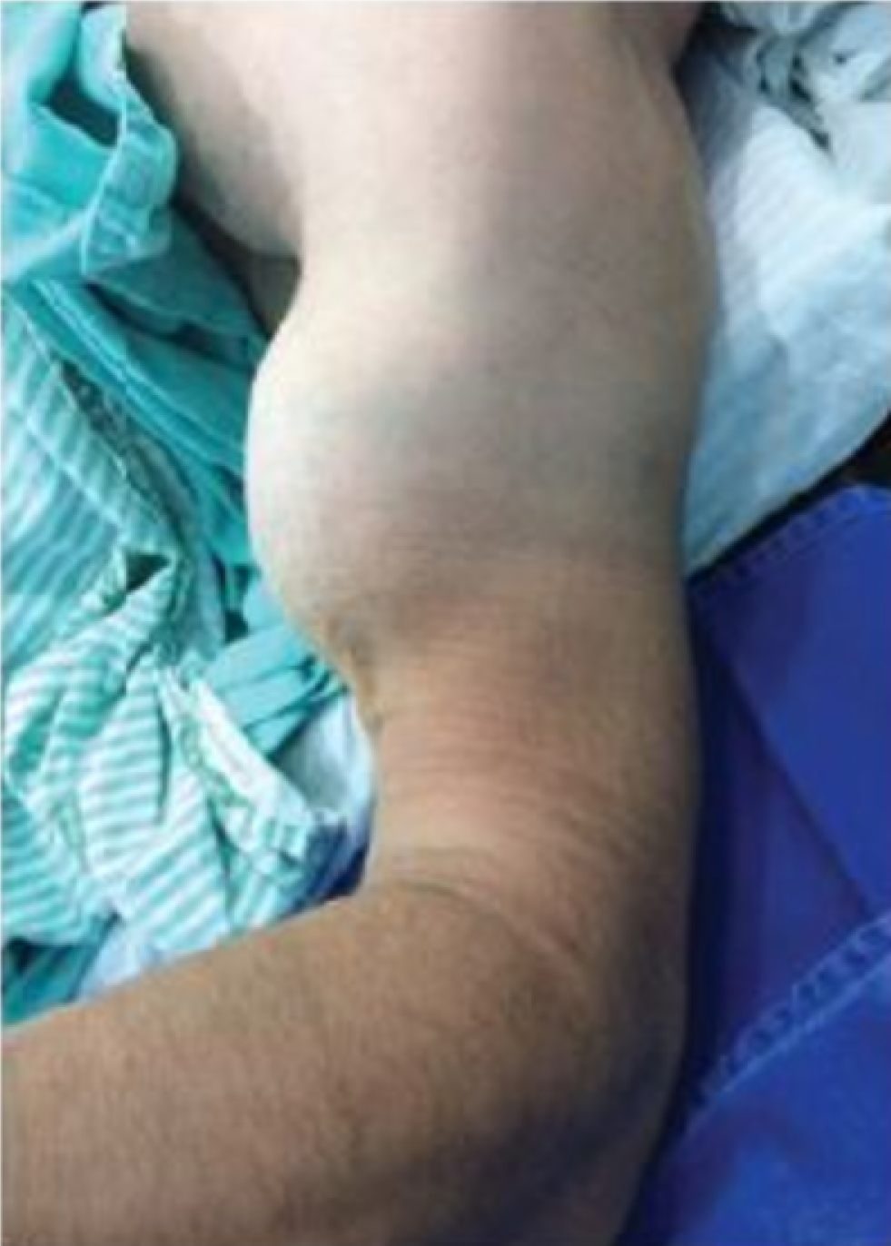
Figure 5 Lateral view showing Popeye deformity from José et al[48].
Citation: José AG, Luís Felipe HFS, Gabriel RSM, Fernando MI. Treatment of the Distal Biceps Brachii Tendon Rupture Using the Three Mini-Incisions Technique: Evaluation through MEPS and DASH. Ortho Rheum Open Access J. 2019; 14: 555888. Copyright© The Authors 2019. Published by Juniper Publishers INC. This work is licensed under Creative Commons Attribution 4.0 License.
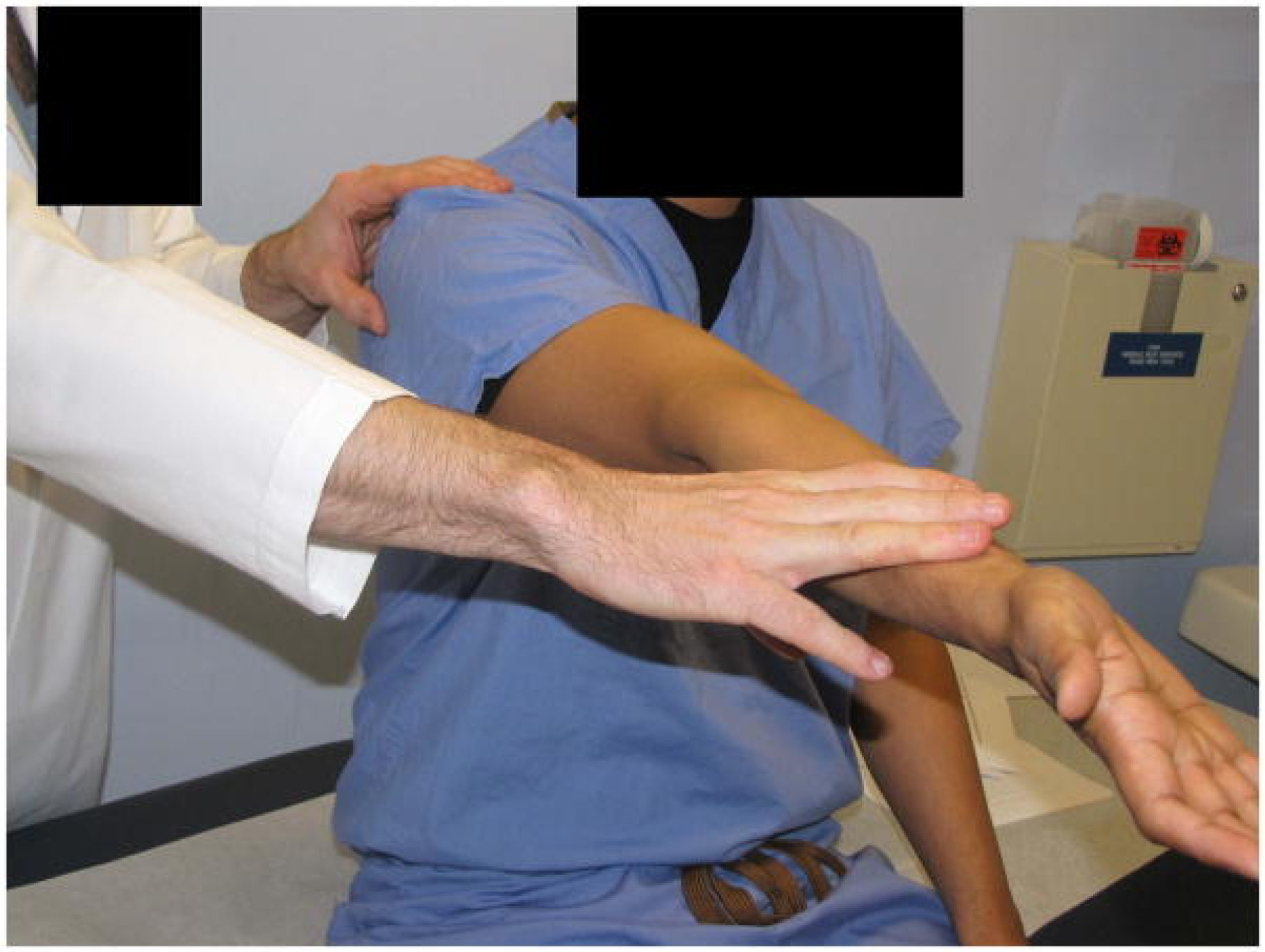
Figure 6 Speed test from Jain et al[47].
Citation: Jain NB, Wilcox RB 3rd, Katz JN, Higgins LD. Clinical examination of the rotator cuff. PM R 2013; 5: 45-56. Copyright© The Authors 2013. Published by John Wiley and Sons. The authors have obtained the permission for figure (Supplementary material).
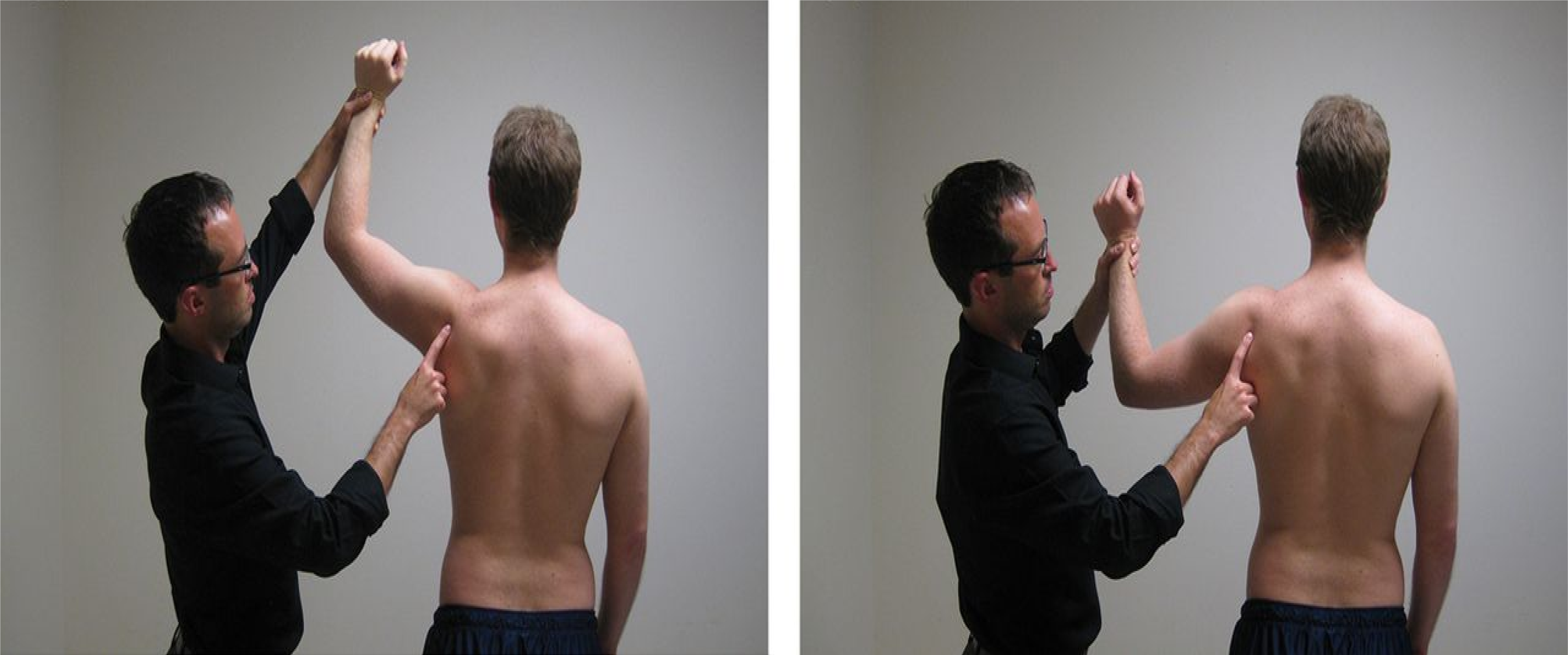
Figure 7 O’Driscoll dynamic labral shear test from Myer et al[57].
Citation: Myer CA, Hegedus EJ, Tarara DT, Myer DM. A user's guide to performance of the best shoulder physical examination tests. Br J Sports Med 2013; 47: 903-907. Copyright© The Authors 2013. Published by BMJ Publishing Group Ltd. The authors have obtained the permission for figure (Supplementary material).
- Citation: Lalehzarian SP, Agarwalla A, Liu JN. Management of proximal biceps tendon pathology. World J Orthop 2022; 13(1): 36-57
- URL: https://www.wjgnet.com/2218-5836/full/v13/i1/36.htm
- DOI: https://dx.doi.org/10.5312/wjo.v13.i1.36


