Copyright
©The Author(s) 2021.
World J Orthop. Jun 18, 2021; 12(6): 423-432
Published online Jun 18, 2021. doi: 10.5312/wjo.v12.i6.423
Published online Jun 18, 2021. doi: 10.5312/wjo.v12.i6.423
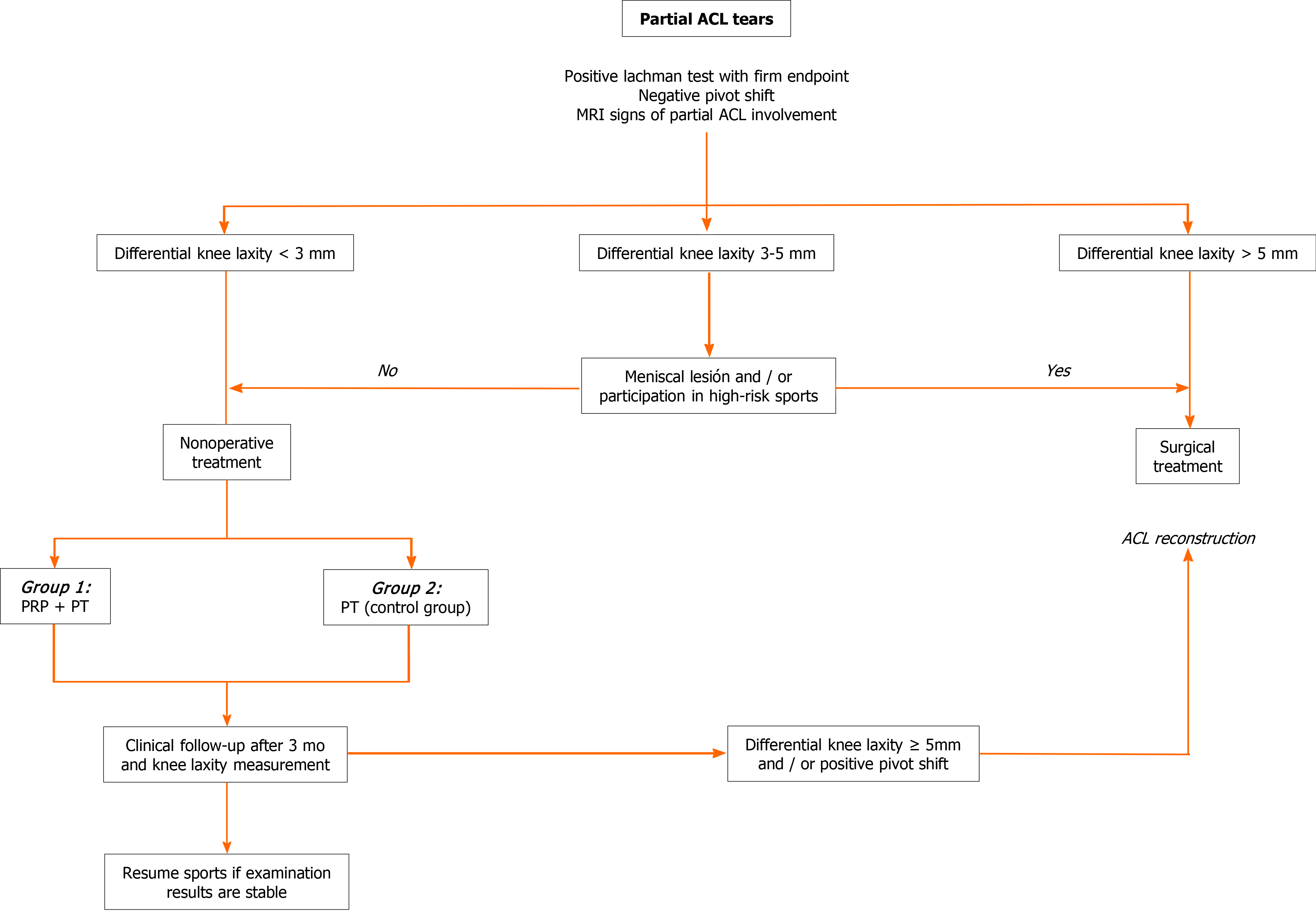
Figure 1 Flow chart for partial anterior cruciate ligament tears.
Management algorithm used to select treatment for partial anterior cruciate ligament tears. ACL: Anterior cruciate ligament; MRI: Magnetic resonance imaging; PRP: Platelet-rich plasma; PT: Physical therapy.
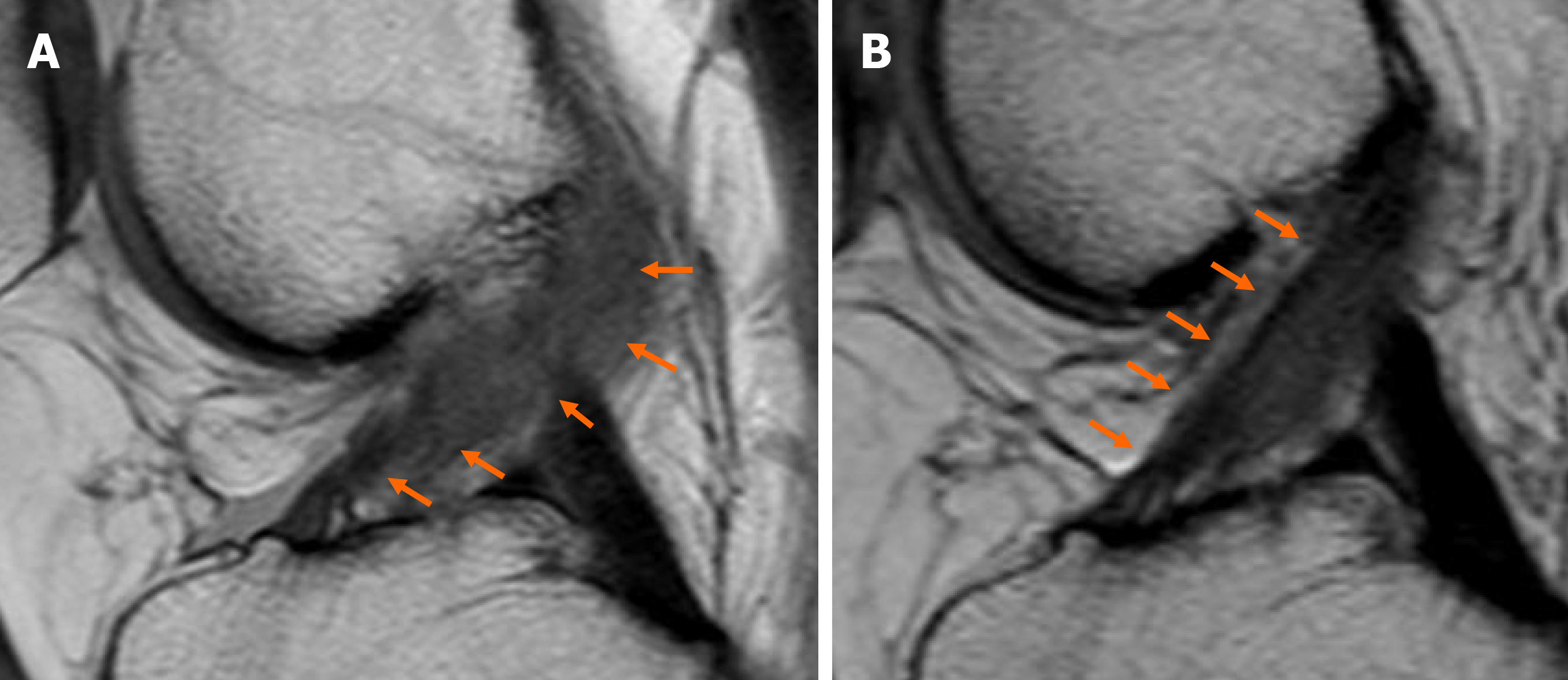
Figure 2 Magnetic resonance images.
A: Baseline magnetic resonance imaging (MRI) showed widening of anterior cruciate ligament (ACL) fibers with continuity of fibers (orange arrows). Total score of 4 points according to Van Meer’s classification; B: Six months after platelet-rich plasma injection, MRI showed an improvement in the signal intensity as well as tension of ACL fibers (orange arrows). Total score of 0 points according to Van Meer’s classification (MRI sequence: sagittal proton density weighted turbo spin echo).
- Citation: Zicaro JP, Garcia-Mansilla I, Zuain A, Yacuzzi C, Costa-Paz M. Has platelet-rich plasma any role in partial tears of the anterior cruciate ligament? Prospective comparative study. World J Orthop 2021; 12(6): 423-432
- URL: https://www.wjgnet.com/2218-5836/full/v12/i6/423.htm
- DOI: https://dx.doi.org/10.5312/wjo.v12.i6.423


