Copyright
©The Author(s) 2021.
World J Orthop. Mar 18, 2021; 12(3): 140-151
Published online Mar 18, 2021. doi: 10.5312/wjo.v12.i3.140
Published online Mar 18, 2021. doi: 10.5312/wjo.v12.i3.140
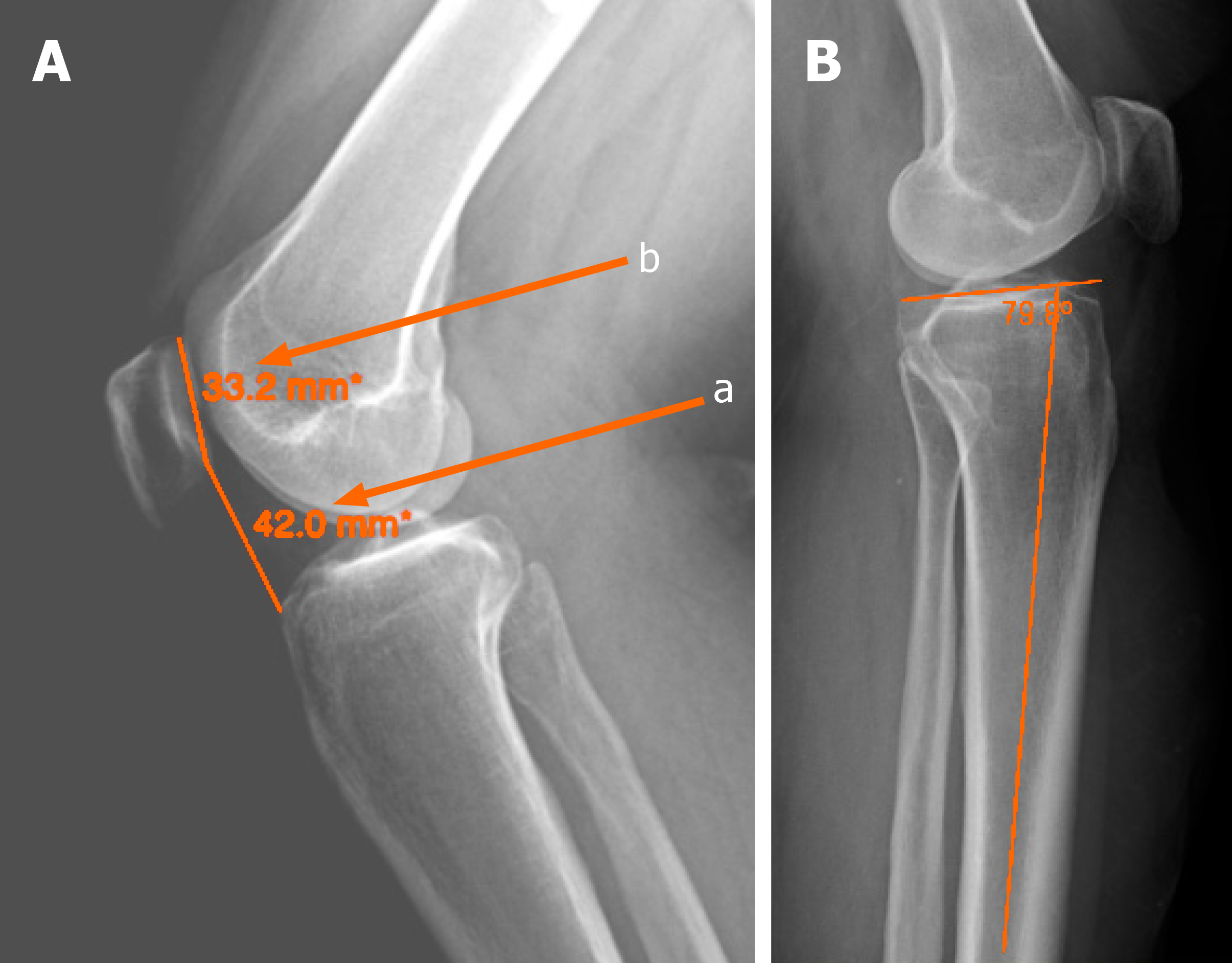
Figure 1 Knee X-ray images.
A: Caton-Deschamps Index (a/b) for evaluation of the patella height; B: Posterior proximal tibial angle.
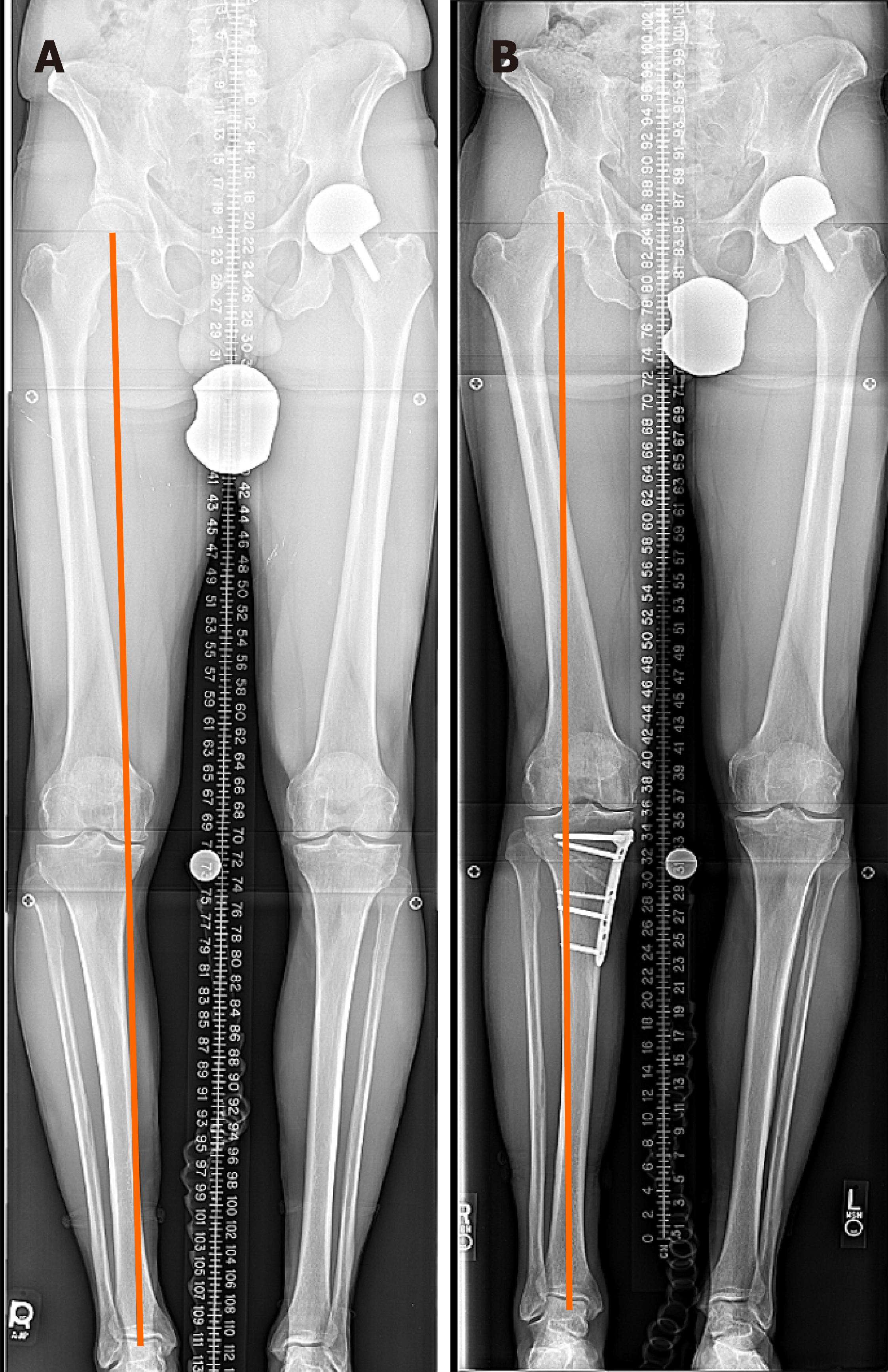
Figure 2 High tibial osteotomy with plate and screws.
A: Pre-operation; B: Post-operation.
Figure 3 High tibial osteotomy with external fixator.
A and B: Before correction medial proximal tibial angle: 82, joint line obliquity angle: 2, mechanical axis deviation: 33 mm, lateral distal femoral angle: 90; C: After correction with external fixator; D: After removal of external fixator.
- Citation: Ghasemi SA, Zhang DT, Fragomen A, Rozbruch SR. Proximal tibial osteotomy for genu varum: Radiological evaluation of deformity correction with a plate vs external fixator. World J Orthop 2021; 12(3): 140-151
- URL: https://www.wjgnet.com/2218-5836/full/v12/i3/140.htm
- DOI: https://dx.doi.org/10.5312/wjo.v12.i3.140


