Copyright
©The Author(s) 2021.
World J Orthop. Nov 18, 2021; 12(11): 938-944
Published online Nov 18, 2021. doi: 10.5312/wjo.v12.i11.938
Published online Nov 18, 2021. doi: 10.5312/wjo.v12.i11.938
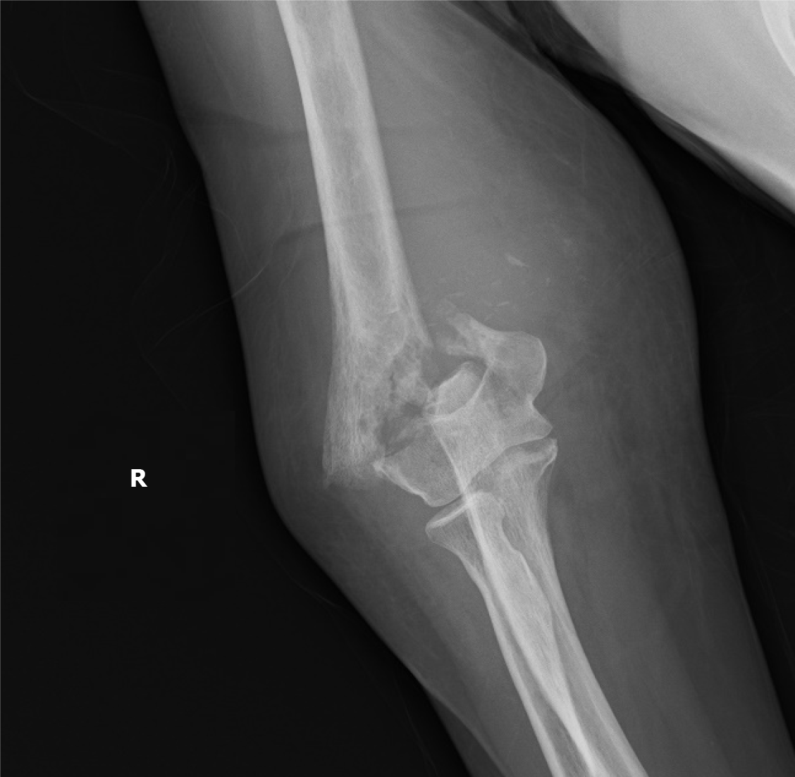
Figure 1
Displaced supracondylar humerus fracture of the right elbow on plain X-ray.
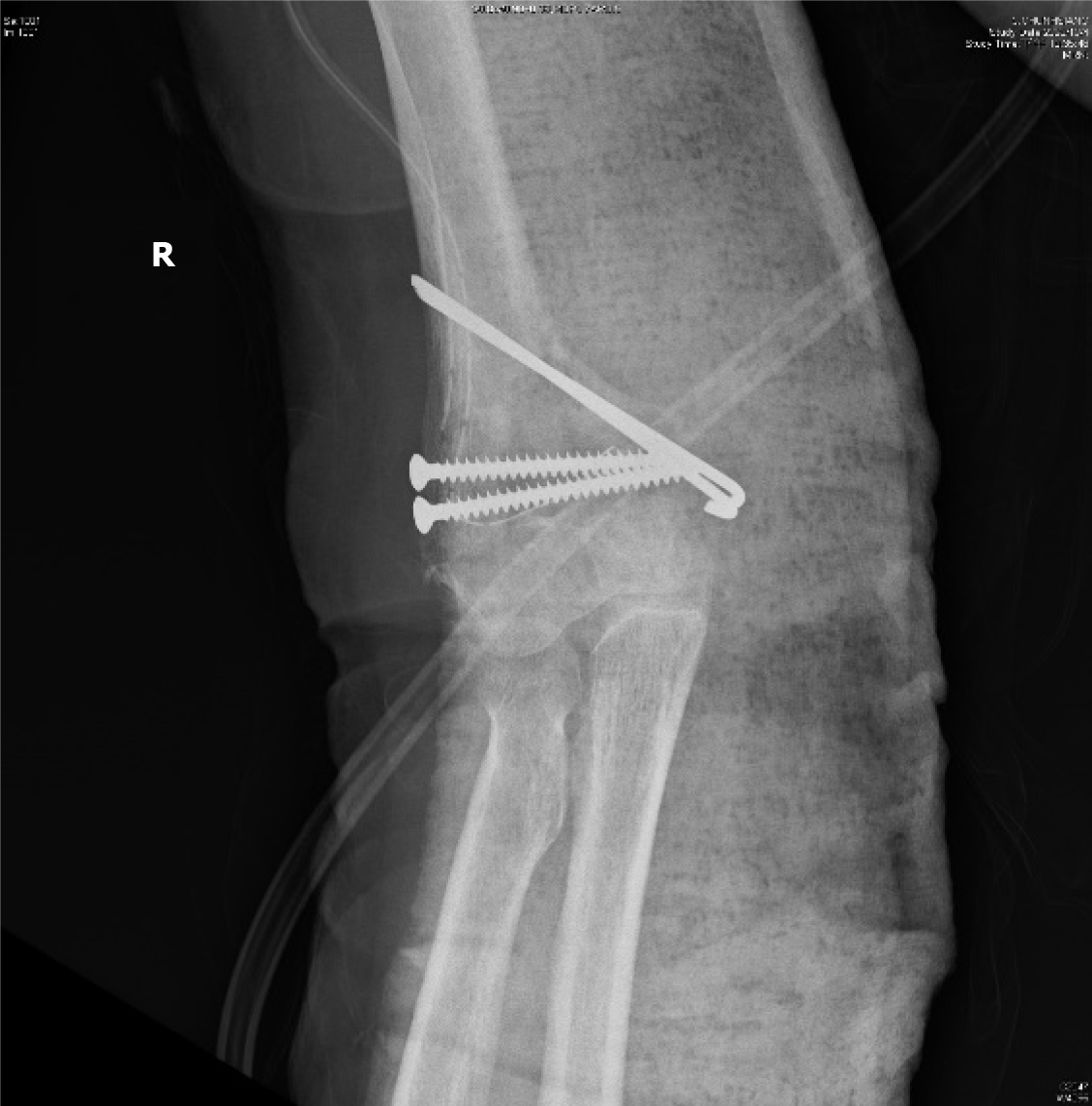
Figure 2
Displaced supracondylar humerus fracture status post-open reduction and internal fixation with screws and Kirschner wires.
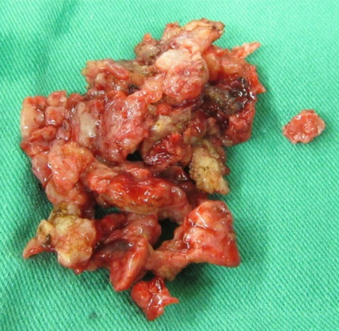
Figure 3
Intraoperative finding of unusual necrotic bone tissues, status post-sequestrectomy.
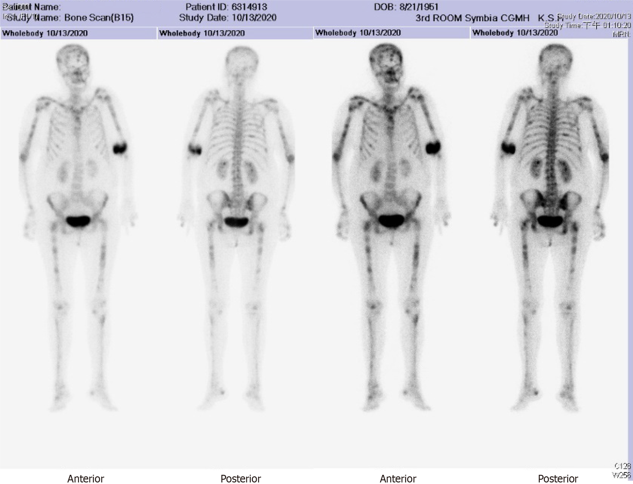
Figure 4 Bone scan revealed multiple uptake.
- Citation: Yang CH, Kuo FC, Lee CH. Pathological humerus fracture due to anti-interferon-gamma autoantibodies: A case report. World J Orthop 2021; 12(11): 938-944
- URL: https://www.wjgnet.com/2218-5836/full/v12/i11/938.htm
- DOI: https://dx.doi.org/10.5312/wjo.v12.i11.938


