Copyright
©The Author(s) 2020.
World J Orthop. Aug 18, 2020; 11(8): 357-363
Published online Aug 18, 2020. doi: 10.5312/wjo.v11.i8.357
Published online Aug 18, 2020. doi: 10.5312/wjo.v11.i8.357
Figure 1 X-ray taken after the initial trauma showing a right avulsion fracture of the apophysis of the anterior inferior iliac spine.
Figure 2 X-ray taken 3 mo after initial trauma showing callus formation in the anterior inferior iliac spine.
Figure 3 X-ray showing a round pedunculated bony protrusion on the superolateral aspect of the right anterior inferior iliac spine.
Figure 4 Imaging examinations.
A: T1-weighted magnetic resonance images showed low signal intensity in the mass; B and C: T2 mDIXON water magnetic resonance images, gadolinium-enhanced T1 mDIXON water images showing high signal intensity around the mass.
Figure 5 Histological section of an osteochondroma showing the three layers.
Red arrow: Perichondrium; Green arrow: Cartilage cap; Yellow arrow: Underlying bone. Hematoxylin and eosin stain, × 100.
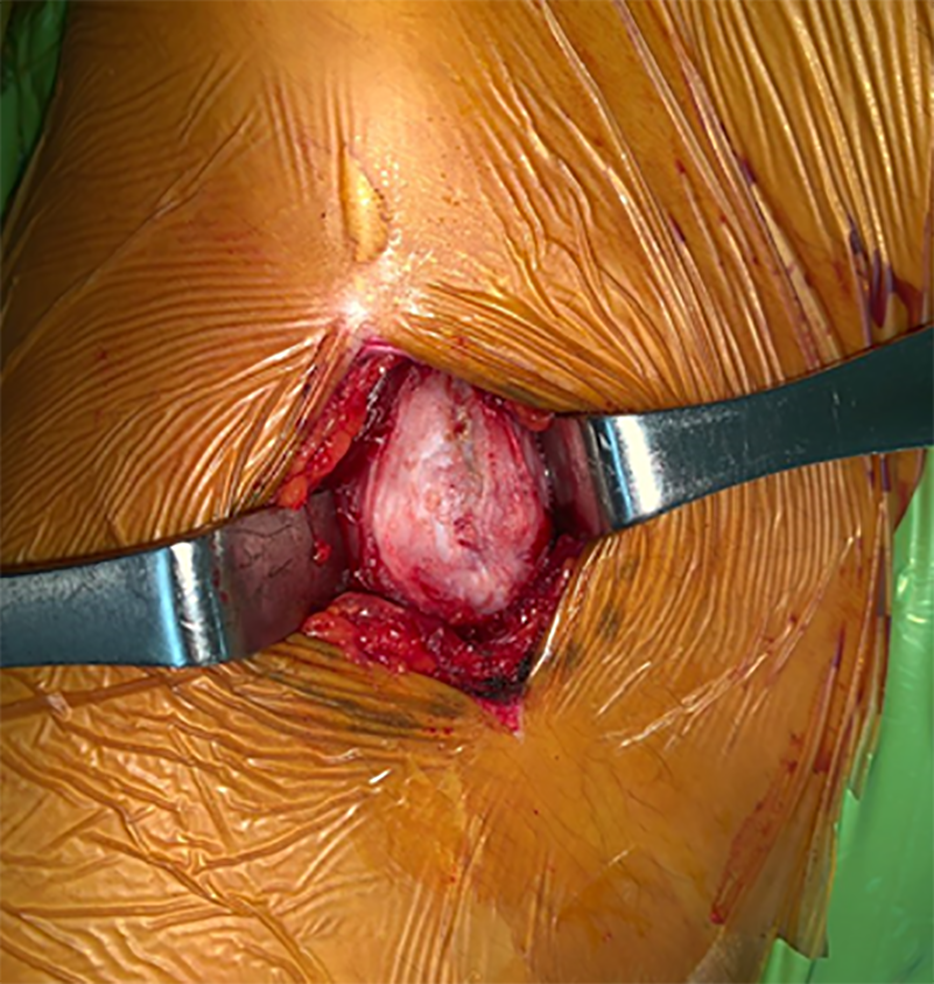
Figure 6 Intraoperatively, an osteochondroma with a smooth margin and cartilaginous cap was found.
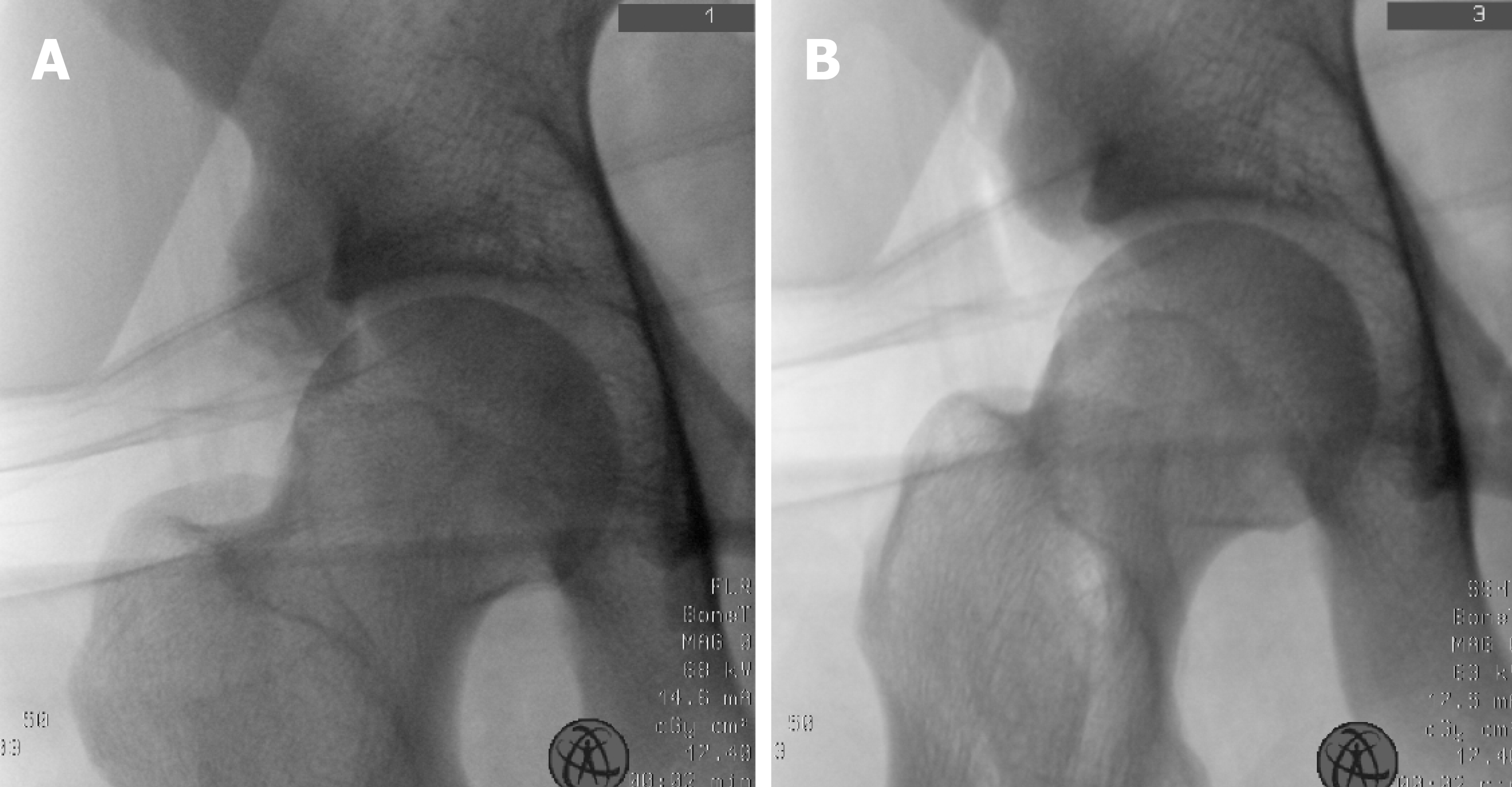
Figure 7 Intraoperative fluoroscopic device showed the bony border being not the same as that of the unaffected side.
A: C-arm image before cutting; B: C-arm image after cutting.
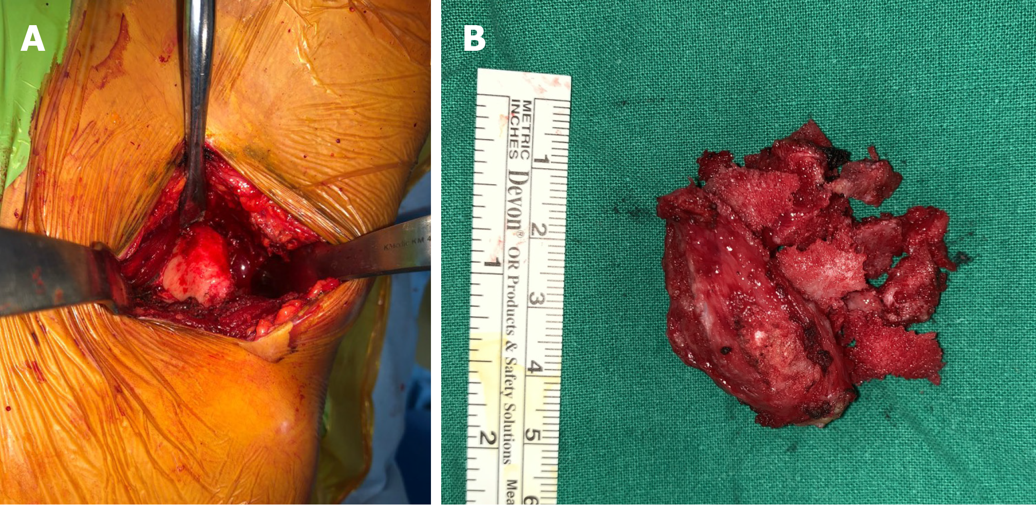
Figure 8 Confirm the removal with additional osteotomy by only the surface being peeled without the osteotome internally proceeding.
A: After excision, the fresh bone was exposed; B: A 4 cm × 4 cm specimen was excised.
Figure 9 Postoperative X-ray that shows the border of the bone being different from that of the unaffected side.
- Citation: Cho HG, Kwon HY, Lee YC, Lee YC, Kweon SH. Osteochondroma formation after avulsion fracture of anterior inferior iliac spine: A case report. World J Orthop 2020; 11(8): 357-363
- URL: https://www.wjgnet.com/2218-5836/full/v11/i8/357.htm
- DOI: https://dx.doi.org/10.5312/wjo.v11.i8.357


