Copyright
©The Author(s) 2020.
World J Orthop. Mar 18, 2020; 11(3): 197-205
Published online Mar 18, 2020. doi: 10.5312/wjo.v11.i3.197
Published online Mar 18, 2020. doi: 10.5312/wjo.v11.i3.197
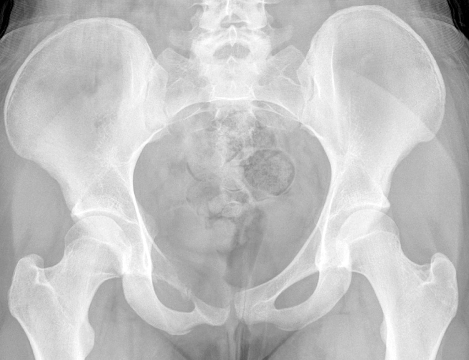
Figure 1 Pre-operative anteroposterior pelvis.
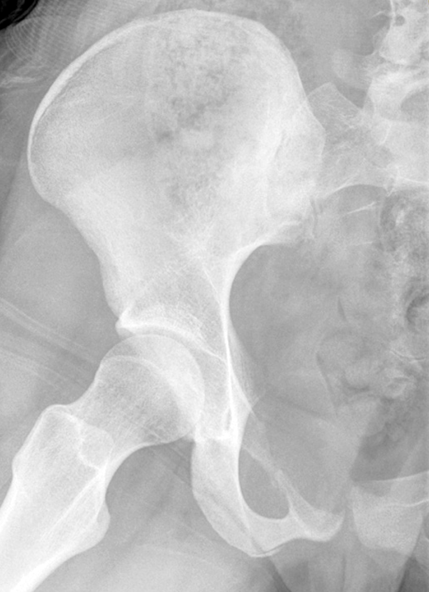
Figure 2 Frog-leg lateral view right hip.
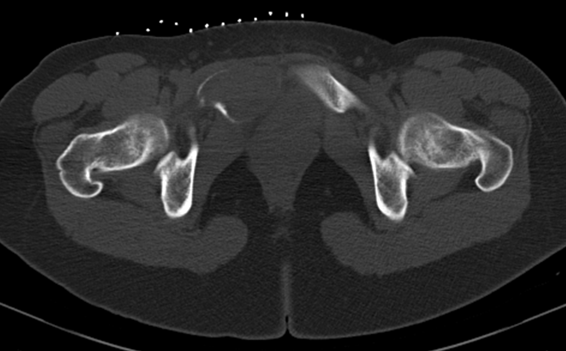
Figure 3 Axial computed tomography pelvis.
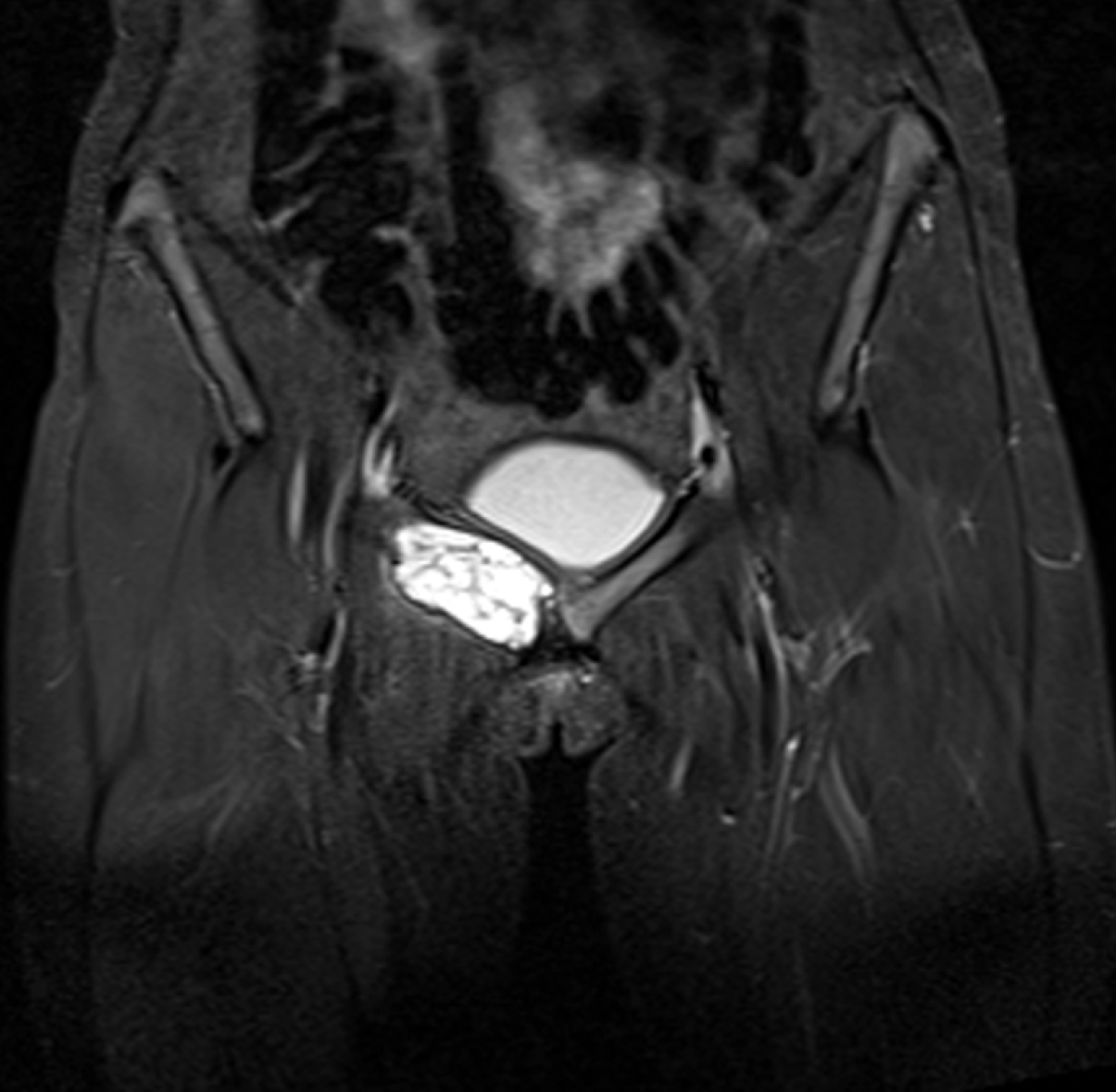
Figure 4 Coronal short tau inversion recovery magnetic resonance imaging.
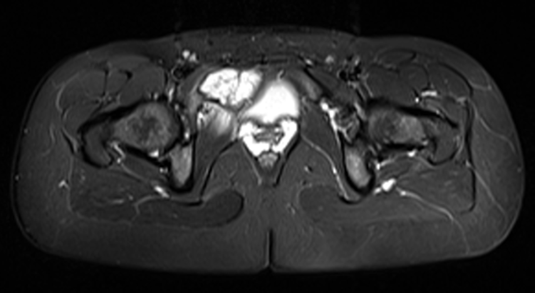
Figure 5 Axial short tau inversion recovery magnetic resonance imaging.
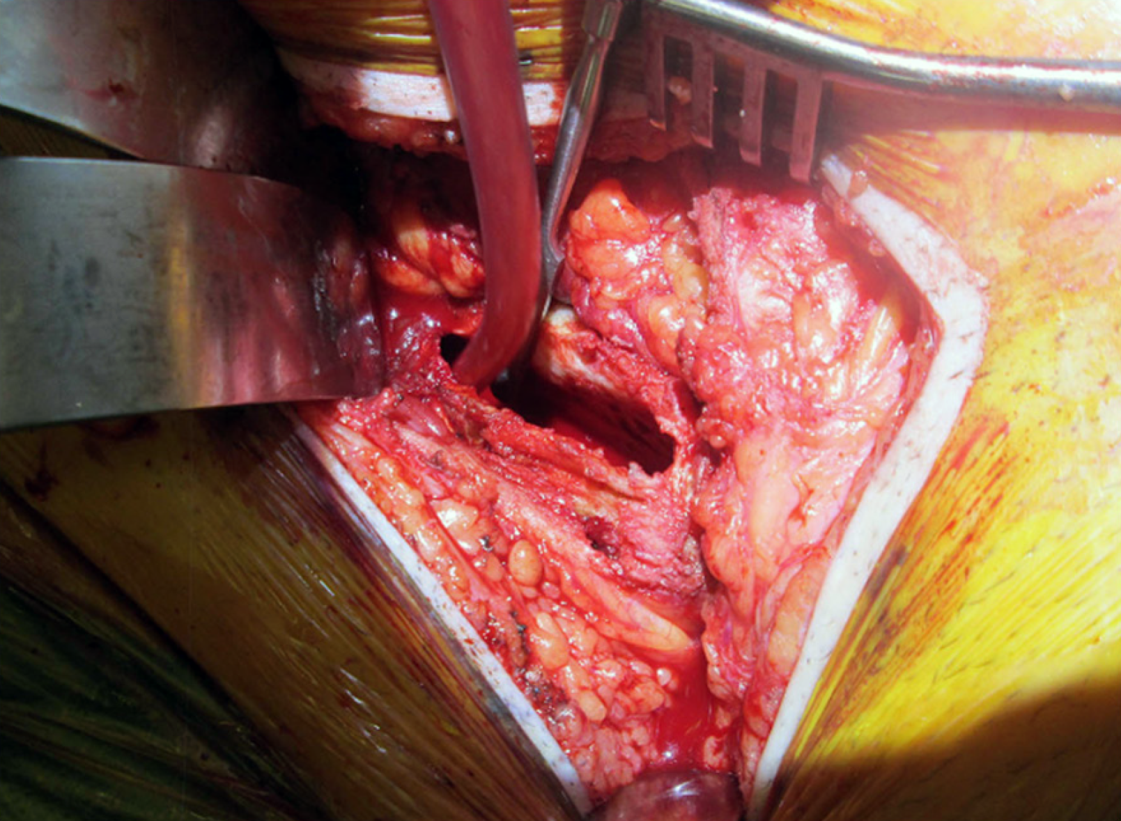
Figure 6 Intra-operative view of lesion.
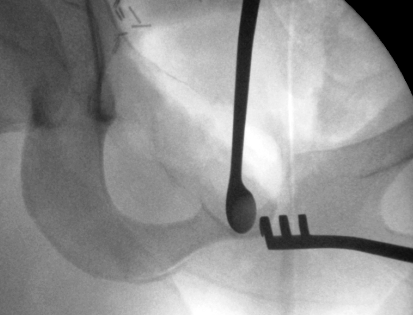
Figure 7 Intra-operative screening demonstrating curettage.
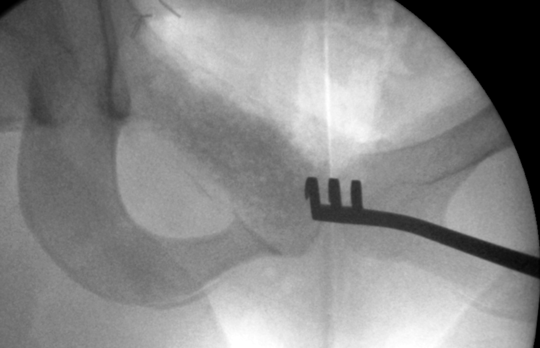
Figure 8 Intra-operative screening with bone graft substitute.
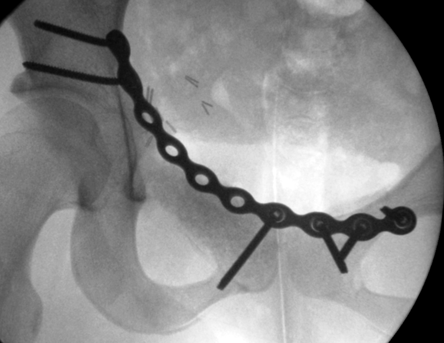
Figure 9 Intra-operative screening with open reduction internal fixation.
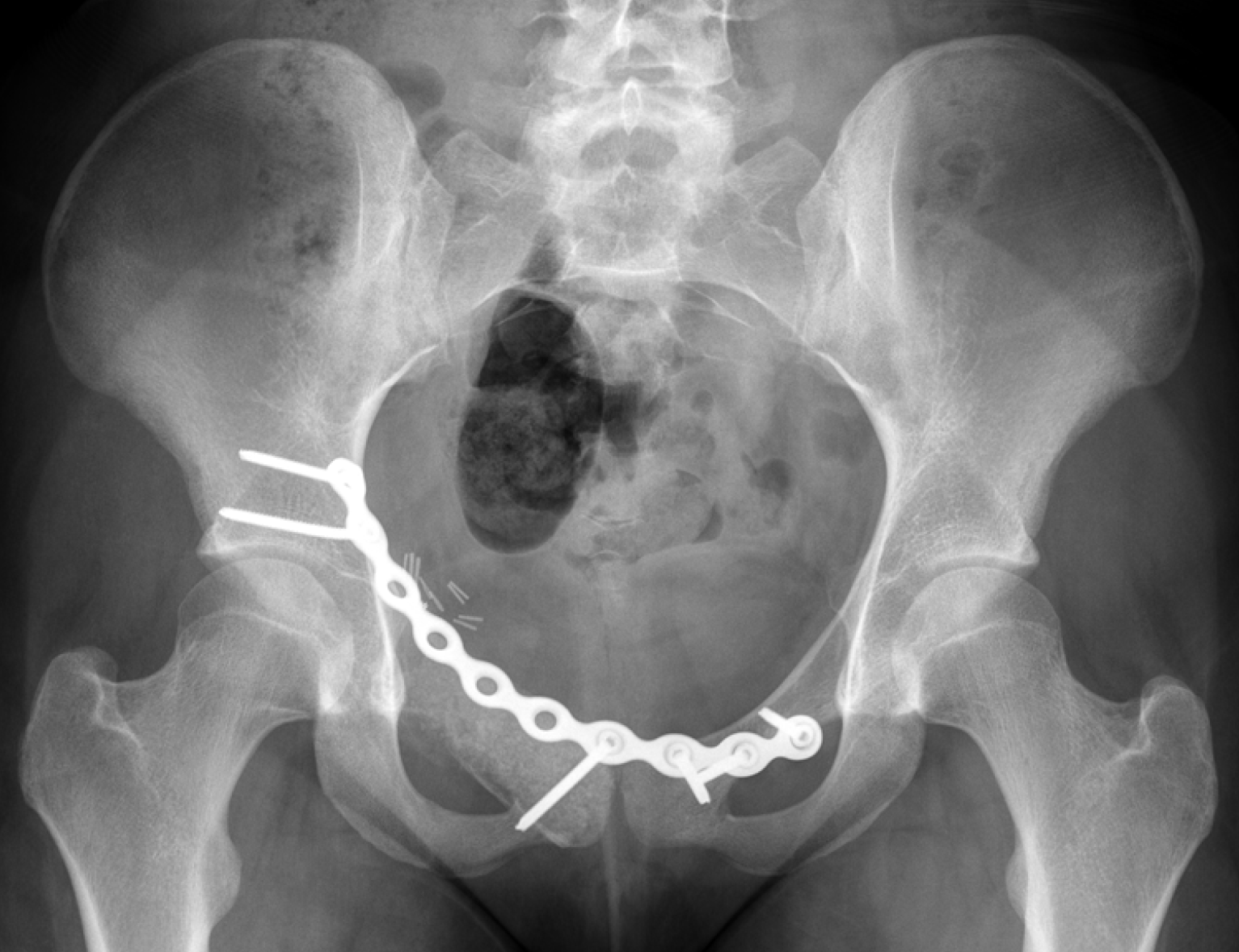
Figure 10 Post-operative X-ray anteroposterior pelvis with open reduction internal fixation and bone graft substitute.
- Citation: Downey C, Daly A, Molloy AP, O’Daly BJ. Atraumatic groin pain secondary to an aneurysmal bone cyst: A case report and literature review. World J Orthop 2020; 11(3): 197-205
- URL: https://www.wjgnet.com/2218-5836/full/v11/i3/197.htm
- DOI: https://dx.doi.org/10.5312/wjo.v11.i3.197


