Copyright
©The Author(s) 2020.
World J Orthop. Nov 18, 2020; 11(11): 516-522
Published online Nov 18, 2020. doi: 10.5312/wjo.v11.i11.516
Published online Nov 18, 2020. doi: 10.5312/wjo.v11.i11.516
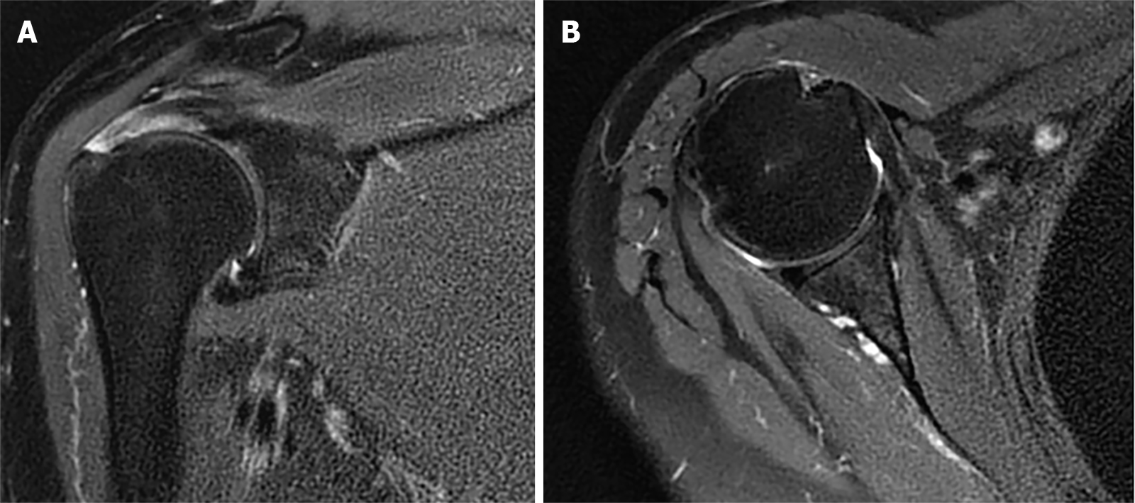
Figure 1 Magnetic resonance imaging of the right shoulder before surgery.
A: The full-thickness rotator cuff tear; B: The intact glenoid of the right shoulder.
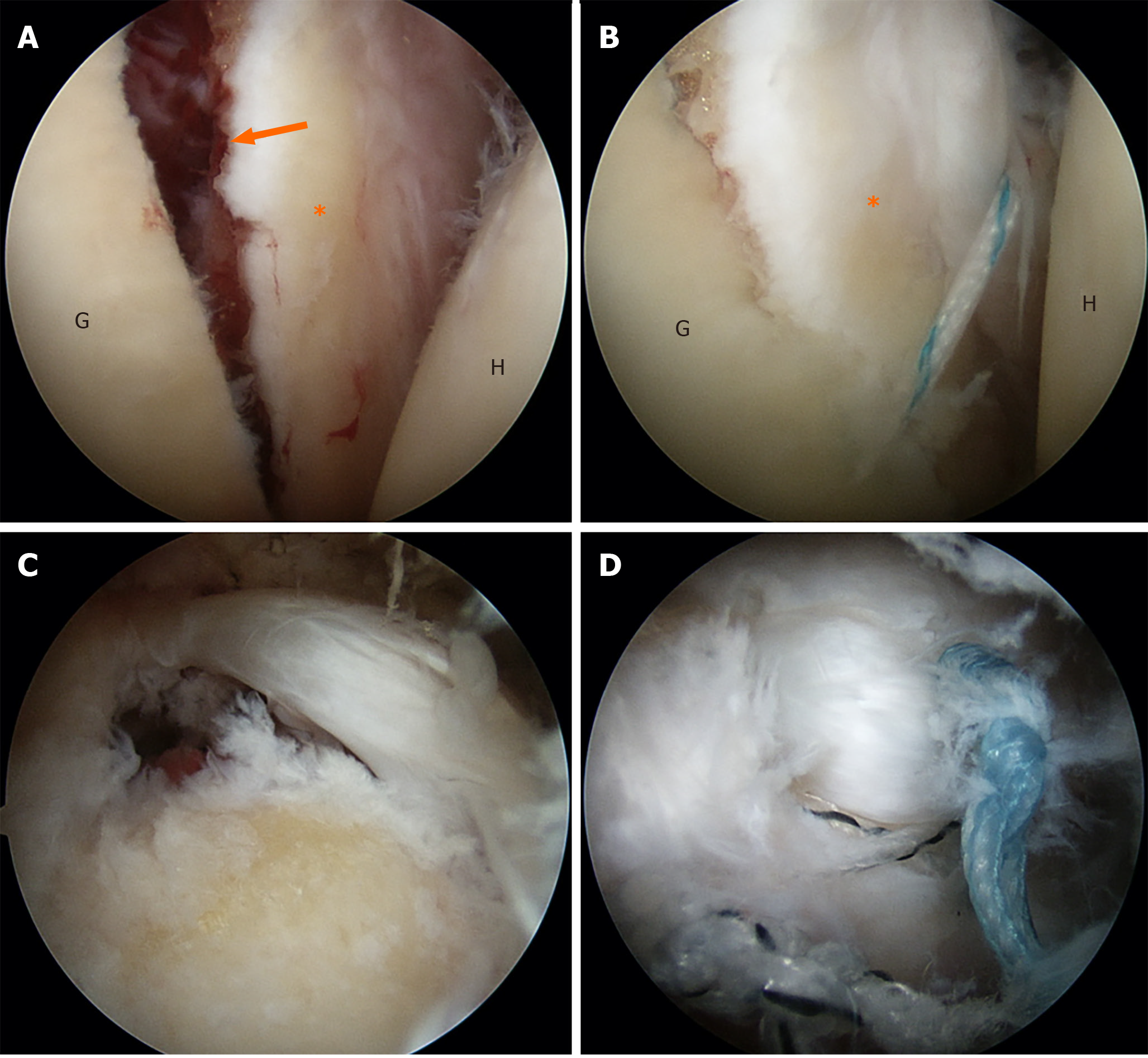
Figure 2 Intraoperative photographs.
A: A fresh fracture with displacement at the anteroinferior glenoid rim. The size of the fragment was about 5 mm and 15 mm in the anterior-posterior and superior-inferior directions, respectively; B: The suture anchors for fixation of the fracture; C: The small-sized full-thickness supraspinatus tear; D: Torn rotator cuff was repaired by the double-row arthroscopic technique. G: Glenoid; H: Humeral head; Star: The fragment of anteroinferior glenoid with intact anterior joint capsule; Black arrow: Fresh fracture line.
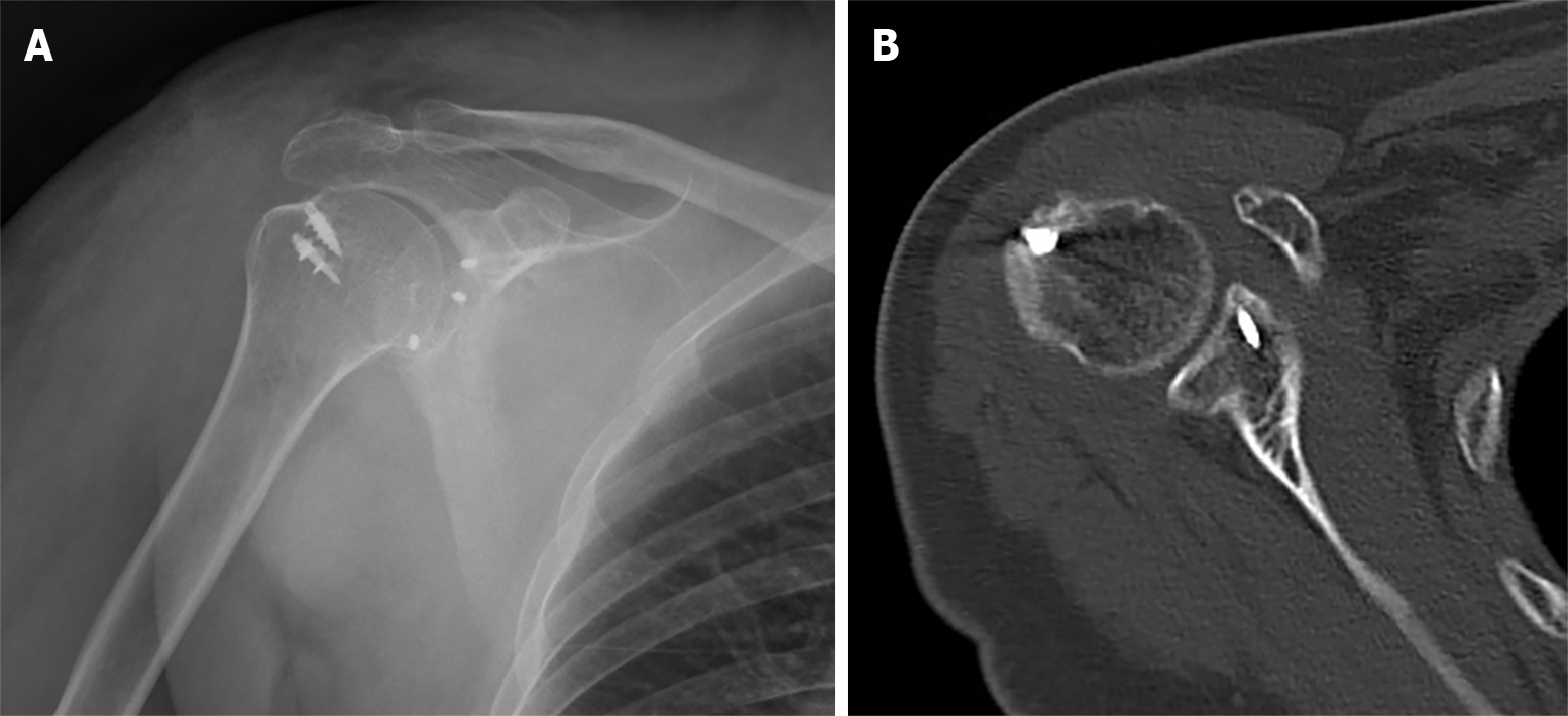
Figure 3 Radiograph at the 3-mo follow-up.
A: The position of the anchors and the computed tomography scan are shown; B: Good healing of the fracture.
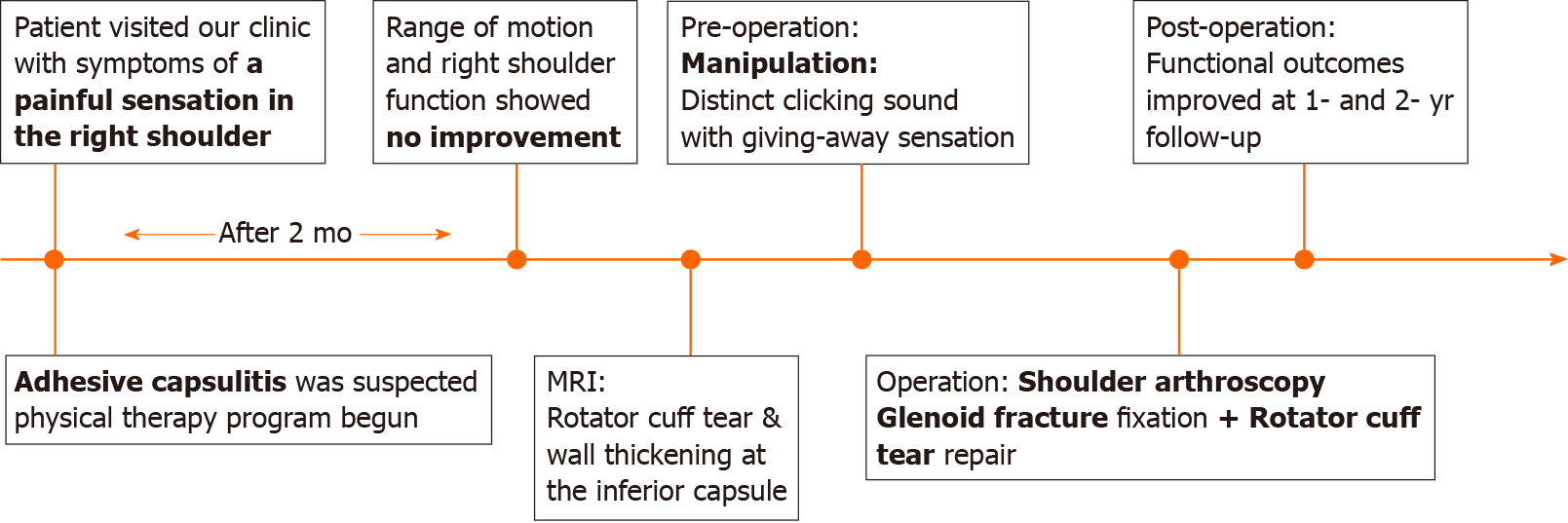
Figure 4 Timeline of the case.
MRI: Magnetic resonance imaging.
- Citation: Chiang CH, Tsai TC, Tung KK, Chih WH, Yeh ML, Su WR. Treatment of a rotator cuff tear combined with iatrogenic glenoid fracture and shoulder instability: A rare case report . World J Orthop 2020; 11(11): 516-522
- URL: https://www.wjgnet.com/2218-5836/full/v11/i11/516.htm
- DOI: https://dx.doi.org/10.5312/wjo.v11.i11.516


