Copyright
©The Author(s) 2020.
World J Orthop. Nov 18, 2020; 11(11): 483-491
Published online Nov 18, 2020. doi: 10.5312/wjo.v11.i11.483
Published online Nov 18, 2020. doi: 10.5312/wjo.v11.i11.483
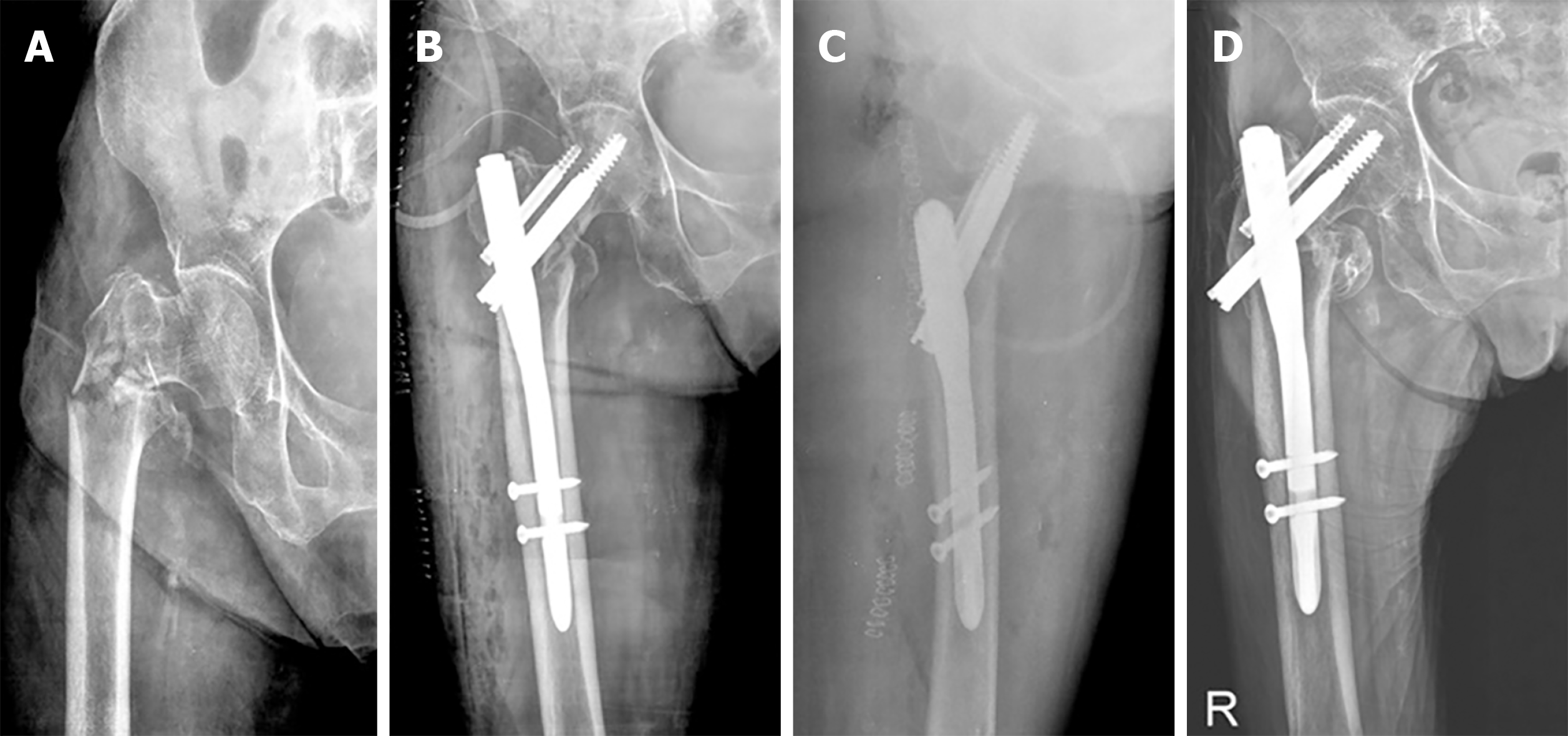
Figure 1 Radiographic results.
A: A preoperative radiograph shows an intertrochanteric fracture with AO classification type A3.2. in a 79-year-old female patient. She underwent closed reduction and fixation with proximal femoral nail. Immediate postoperative radiographs demonstrate good reduction, tip-apex distance of 12.1 mm and Cleveland Index of 5 on anteroposterior (B) and axial view (C). D: A radiograph taken at follow-up duration of 3 years shows bone union with the sliding distance of 14.5 mm.
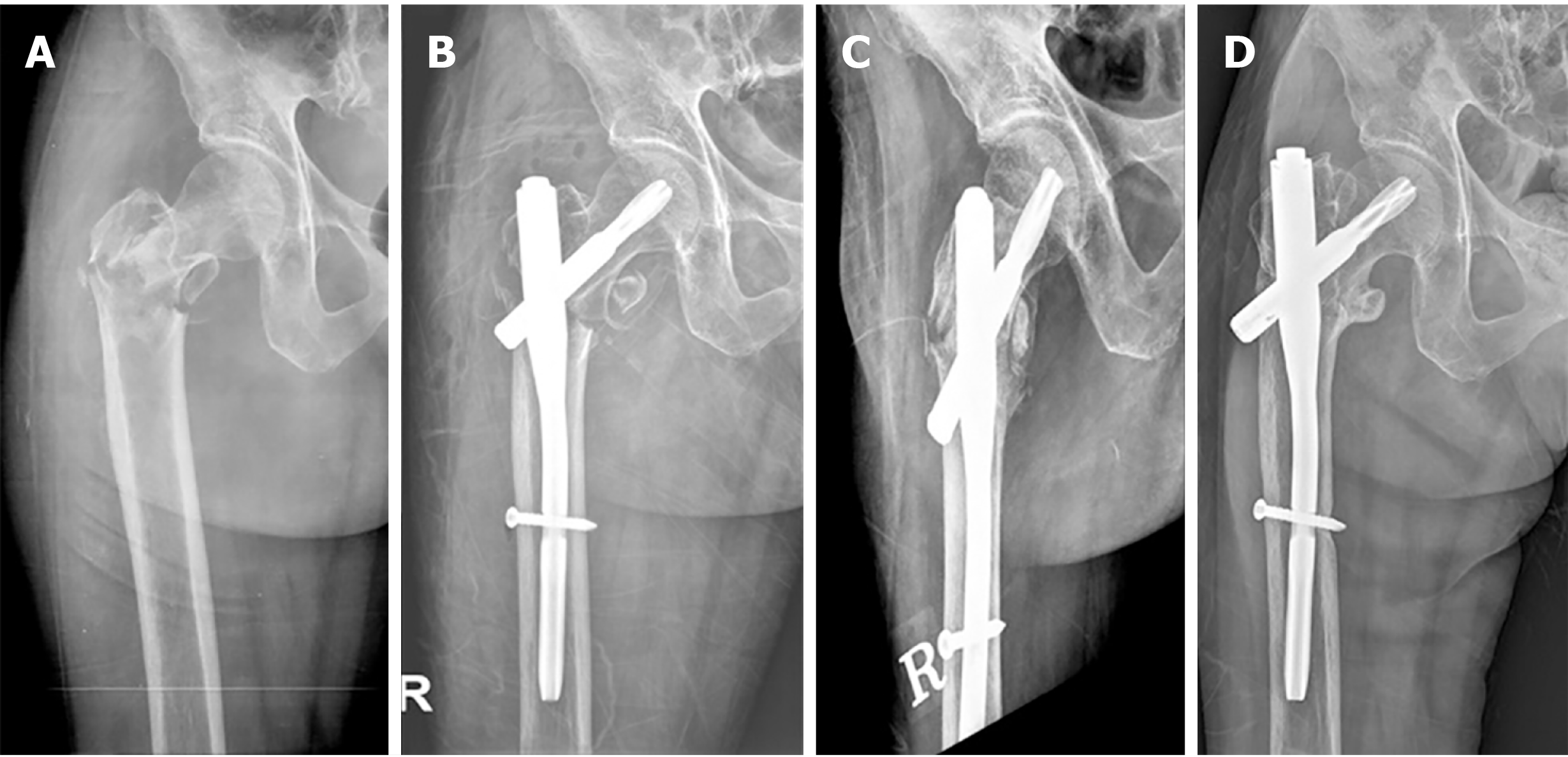
Figure 2 Radiographic results.
A: A preoperative radiograph shows an intertrochanteric fracture with AO classification type A3.3 in a 59-year-old female patient. She underwent closed reduction and fixation with proximal femoral nail antirotation. B and C: Immediate postoperative radiographs demonstrate distal fragment is distracted, leaving a fracture gap, tip-apex distance of 21.2 mm and Cleveland Index of 5 on anteroposterior (B) and axial view (C). D: A radiograph taken at follow-up duration of 7 years shows bone union with the sliding distance of 8.6 mm.
- Citation: Baek SH, Baek S, Won H, Yoon JW, Jung CH, Kim SY. Does proximal femoral nail antirotation achieve better outcome than previous-generation proximal femoral nail? World J Orthop 2020; 11(11): 483-491
- URL: https://www.wjgnet.com/2218-5836/full/v11/i11/483.htm
- DOI: https://dx.doi.org/10.5312/wjo.v11.i11.483


