Copyright
©The Author(s) 2019.
World J Orthop. Aug 18, 2019; 10(8): 304-309
Published online Aug 18, 2019. doi: 10.5312/wjo.v10.i8.304
Published online Aug 18, 2019. doi: 10.5312/wjo.v10.i8.304
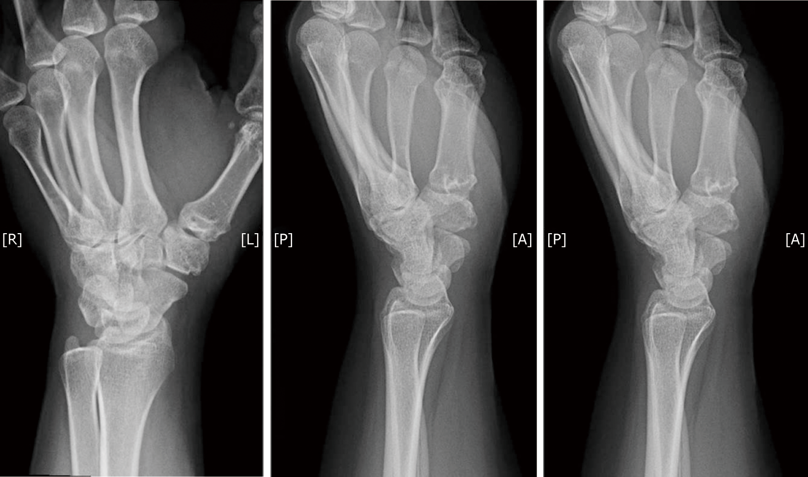
Figure 1 X-ray of patients’ left hand in three planes showing signs of basal joint arthritis of her thumb.
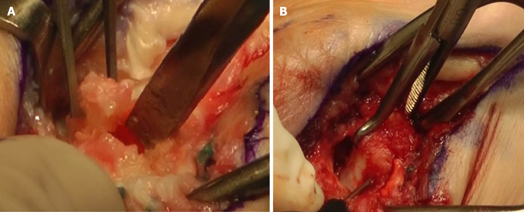
Figure 2 Intraoperative images of the osteotomy.
A: Osteotomy indicated with the chisel after partial trapeziectomy; B: Indicating bone reduction clamp and screw insertion from distal to proximal.
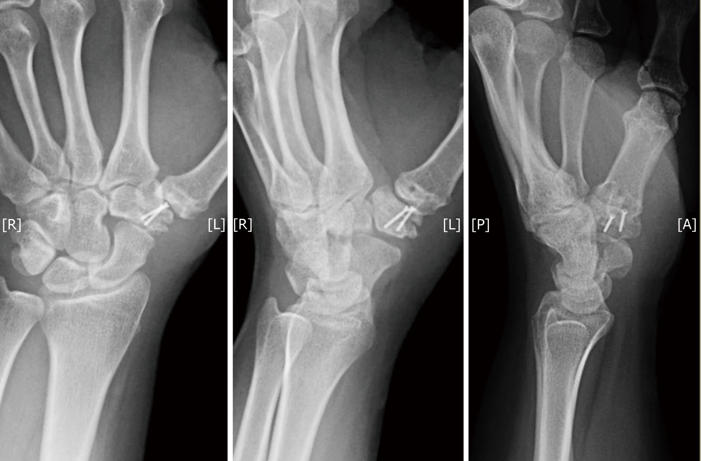
Figure 3 Postoperative x-rays after subtraction hemi-arthroplasty.
- Citation: Bäcker HC, Freibott CE, Rosenwasser MP. Subtraction hemiarthroplasty in basal joint arthritis: A case report. World J Orthop 2019; 10(8): 304-309
- URL: https://www.wjgnet.com/2218-5836/full/v10/i8/304.htm
- DOI: https://dx.doi.org/10.5312/wjo.v10.i8.304


